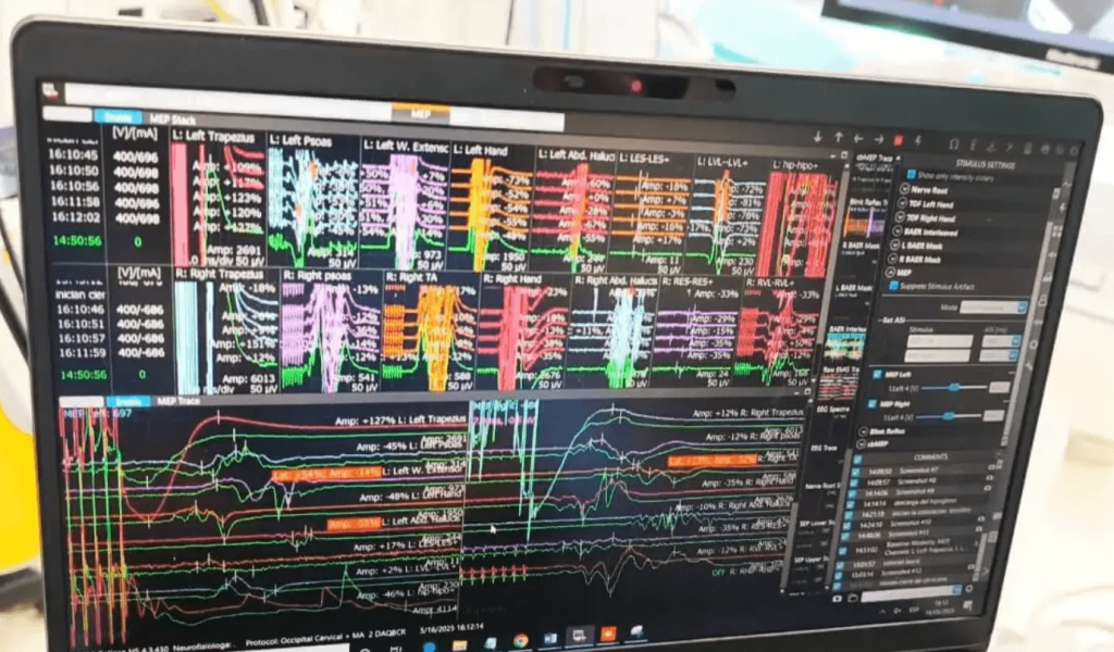
One of the biggest concerns before spine surgery is easy to understand: “what if a nerve or the spinal cord is damaged during the operation?”. Intraoperative neurophysiological monitoring, also known as IONM by its initials in English, helps monitor the function of the spinal cord, the nerve roots and other neurological structures while the surgeon works.
It is not an absolute guarantee nor does it turn a complex surgery into a risk-free procedure. Its value lies elsewhere: it provides real-time information so the team can detect changes, interpret what might be happening and act before an alteration becomes irreversible.
- Intraoperative neurophysiological monitoring records signals from the nervous system during surgery.
- It can monitor motor pathways, sensory pathways and nerve roots using techniques such as motor evoked potentials, somatosensory evoked potentials and electromyography.
- It is especially useful in surgeries with high neurological risk, such as deformity corrections, tumor resections, thoracic surgeries, complex revisions and some lateral approaches.
- It does not eliminate risk 100%, but it can help detect problems in time.
- Anesthesia, blood pressure, temperature, patient positioning and the surgical technique can affect the signals.
- The patient should understand what it provides, what it does not provide and in which cases it makes more sense.
What intraoperative neurophysiological monitoring is
Intraoperative neurophysiological monitoring is a set of techniques that record the electrical activity of the nervous system during an operation. In spine surgery, its main objective is to monitor whether the spinal cord, the nerve roots or certain nerves continue to function properly while the procedure is performed.
Put simply: before starting the most delicate steps, the team obtains baseline signals. During the surgery, those signals are compared with the initial ones. If they change significantly, the neurophysiologist notifies the surgical and anesthesia team to investigate the cause.
The change can be due to many reasons: traction on a nervous structure, compression, altered blood supply, patient positioning, changes in blood pressure, temperature, anesthesia, or a technical problem with the recording. That is why monitoring is not just “watching a screen”; it requires interpretation, communication and a coordinated response.
Why it matters in spine surgery
The spine protects the spinal cord and the nerve roots. In some operations, these structures are close to the surgical field or can be affected by corrective maneuvers, implant placement, decompression, tumor resection or complex approaches.
Severe neurological injury is uncommon, but it can have significant consequences: loss of strength, altered sensation, neuropathic pain, walking difficulties or, in exceptional cases, paralysis. Therefore, in selected surgeries, having a tool that alerts to neurological changes during the procedure can be very valuable.
Monitoring helps move from surgery based only on visible anatomy and imaging to surgery with functional information. In other words, not only is it seen where the structures are, but also how they respond.
Which structures are monitored
The structure of greatest concern in many surgeries is the spinal cord, especially in cervical and thoracic operations. The cord transmits signals between the brain and the rest of the body. A significant alteration can affect movement or sensation.
Nerve roots can also be monitored; these are the nerves that exit the spine toward the arms, trunk or legs. This can be important in lumbar, cervical or lateral approach surgeries where the roots are close to the working area.
In some cases specific peripheral nerves, motor function of muscle groups or sensory pathways are monitored. The choice depends on the surgery, the spinal level, prior symptoms and the expected neurological risk.
Which techniques are used
Motor evoked potentials
Motor evoked potentials assess the pathway that carries the movement command from the brain to the muscles. They are useful for monitoring motor function, especially when there is risk to the spinal cord. If the motor signal drops significantly, the team must rule out anesthetic, circulatory, technical or surgical causes.
Somatosensory evoked potentials
Somatosensory evoked potentials evaluate pathways related to sensation. They are usually obtained by stimulating peripheral nerves and recording the response along the nervous system. They are particularly useful for detecting changes in sensory conduction.
Spontaneous and triggered electromyography
Electromyography, or EMG, records muscle activity related to roots or nerves. It can help detect irritation of a nerve root or check the proximity of certain instruments or implants to neural structures.
Multimodal monitoring
In many surgeries a single technique is not used, but a combination. This is called multimodal monitoring. It makes sense because each technique looks at a different part of the nervous system. One pathway may remain stable while another changes, and that information helps interpret the situation better.
In which surgeries it is usually most important
Neurophysiological monitoring does not have the same usefulness in all surgeries. In small, low-risk procedures it may not be necessary. Conversely, it usually makes more sense when the spinal cord or nerve roots are at real risk.
Some examples are spinal deformity surgeries, such as complex scoliosis or kyphosis corrections; thoracic surgeries, because the spinal cord has particular relevance there; cervical surgeries with myelopathy or cord compression; revision surgeries, where there may be scar tissue, altered anatomy or previous implants; and some lateral lumbar approaches where the trajectory approaches the lumbar plexus.
It can also be considered in intramedullary or extramedullary tumors, malformations, trauma, large alignment corrections or procedures that foresee manipulation near critical neurological structures.
The decision should not be made out of fashion or marketing. It should be based on the type of surgery, the spinal level, the pathology, the individual risk and the availability of a trained team.
What happens if a signal changes
A signal change does not automatically mean an irreversible injury has occurred. It means one must stop, interpret and act. The response depends on the situation.
The team can first check for a technical problem: displaced electrodes, interference, changes in recording parameters or poor signal quality. Then anesthetic and physiological factors are reviewed: blood pressure, oxygenation, temperature, depth of anesthesia, muscle relaxants or blood loss.
If a surgical cause is suspected, a correction can be loosened, an implant removed or revised, an area of pressure released, the patient’s position modified, irrigation applied, time allowed for signal recovery or an intraoperative imaging test performed if available.
The key is communication. Monitoring is only useful if the information reaches those who can make decisions in a clear and timely manner.
Benefits versus risks and limitations
Potential benefits
The main benefit is detecting neurological changes during the operation, when there may still be time to intervene. It also helps guide decisions: continue, pause, modify a maneuver, review a screw, raise blood pressure or correct an anesthetic factor.
In complex surgeries, it can provide an additional layer of safety and functional documentation. It also forces more careful planning among surgery, anesthesia and neurophysiology.
Limitations
It does not detect all possible injuries. No technique is perfect. There can be false positives, when the signal changes without a real injury, and false negatives, when an injury is not detected but later becomes clinically apparent.
Anesthesia can alter the signals. That is why, when monitoring is used, the anesthetist usually adapts the drugs to preserve reliable recordings. Body temperature, blood pressure, oxygenation, anemia, positioning and prior neurological status also influence results.
Risks and discomforts
Monitoring usually involves placing small electrodes on the scalp, arms, legs or specific muscles. It can leave small marks, local irritation or mild bruising. In stimulation-based techniques, muscle movements can occur during the surgery, controlled within the anesthetic environment.
The main risks do not come so much from the monitoring itself, but from misinterpreting a signal, communicating a change late or using it out of context. That is why it is essential that trained personnel perform it and that the team has a clear response protocol.
Questions to ask before surgery
If spine surgery has been proposed to you, you can ask whether neurophysiological monitoring will be used in your case and why. The answer should be specific, not generic.
Some useful questions are: which structures will be monitored, which techniques will be used, who will interpret the signals, how it is coordinated with anesthesia, what the team will do if an alert appears and whether monitoring will change the surgical plan.
It is also reasonable to ask whether your surgery carries risk to the spinal cord or nerve roots, whether there are personal factors that increase that risk and what other safety measures will be used: preoperative planning, intraoperative imaging, navigation, blood pressure control, bleeding prevention and postoperative neurological checks.
Realistic recovery times
Neurophysiological monitoring does not by itself speed up recovery. Its main role is inside the operating room. Recovery will depend on the surgery performed, the number of levels treated, age, preoperative physical condition, whether there was a neurological deficit before surgery and whether it is a primary surgery or a revision.
After a minor surgery, mobilization can begin the same day or the following day, according to medical advice. In more complex surgeries, such as deformity corrections or extensive revisions, recovery can require weeks or months, with physiotherapy and close follow-up.
If there was preoperative loss of strength, tingling or gait disturbance, neurological recovery can be slow. Sometimes it improves quickly; other times it takes months; and in some cases it does not fully recover if the nerve or spinal cord had been compressed for a long time.
When to go to the emergency room after spine surgery
After spine surgery, you should seek urgent care if you experience new or progressive loss of strength, worsening difficulty walking, loss of bladder or bowel control, numbness in the genital or perineal area, high fever with severe back pain, pain that does not improve with prescribed medication, heavy drainage of fluid or pus from the wound, shortness of breath or chest pain.
These symptoms do not always mean a serious complication, but they should be assessed quickly to rule out hematoma, infection, neurological compression, thrombosis or other postoperative problems.
Myths and realities
Myth: if there is monitoring, neurological injury cannot occur
Reality: monitoring reduces uncertainty and can help detect problems, but it does not eliminate risk 100%.
Myth: it is only used in dangerous surgeries
Reality: it is used when the balance between risk, complexity and usefulness justifies it. It does not mean the surgery will go wrong.
Myth: the machine decides what the surgeon does
Reality: monitoring informs. Decisions are made by the surgical and anesthesia team according to the full context.
Myth: if a signal changes, the damage is already done
Reality: many alerts are reversible if identified and corrected in time.
Myth: all spine surgeries should have monitoring
Reality: it does not always add value. In low-risk surgeries it may not be necessary.
Frequently asked questions
What exactly is intraoperative neurophysiological monitoring?
It is a set of techniques that record signals from the nervous system during surgery. In the spine, it helps monitor the spinal cord, the nerve roots and some motor or sensory pathways while operating.
Does monitoring hurt?
You should not feel pain during the surgery because it is performed under anesthesia. Afterwards, there may be small marks or mild discomfort where electrodes were placed.
Can it prevent paralysis?
It can help detect changes that could precede a neurological injury and allow a rapid response. But it does not guarantee that a complication is impossible.
Is it used in all spine surgeries?
No. It is mainly used when there is relevant risk to the cord or nerves, such as deformities, thoracic surgeries, complex revisions, tumors, myelopathy or some lateral approaches.
What happens if signals are lost during the operation?
The team reviews technical, anesthetic, circulatory and surgical causes. They may change the position, adjust blood pressure, review implants, pause a maneuver or change the plan if necessary.
Does anesthesia affect monitoring?
Yes. Some drugs can modify the signals. That is why anesthesia and neurophysiology must coordinate to obtain reliable recordings.
Who looks at the signals?
They are normally recorded and interpreted by personnel specialized in intraoperative neurophysiology, in communication with the surgeon and the anesthetist.
Does monitoring replace the surgeon’s experience?
No. It is a support tool. Safety depends on correct indication, planning, surgical technique, anesthesia, communication and postoperative follow-up.
Can there be false alarms?
Yes. A signal can change due to anesthesia, temperature, blood pressure, electrodes or interference. That is why interpretation must be careful.
What should I ask before my operation?
Ask whether your surgery has neurological risk, whether monitoring will be used, which pathways will be monitored, who will interpret the signals and how the team will act if an alert appears.
Glossary
- Intraoperative neurophysiological monitoring: recording of nervous system function during surgery.
- IONM: initials in English for intraoperative neurophysiological monitoring.
- Motor evoked potentials: technique that evaluates the motor pathway from the brain to the muscles.
- Somatosensory evoked potentials: technique that evaluates sensory pathways.
- Electromyography: recording of electrical muscle activity related to nerves or roots.
- Nerve root: structure that exits the cord or the dural sac toward the arms, trunk or legs.
- Spinal cord: the main communication pathway between brain and body.
- Intraoperative alert: a relevant change in a signal that requires the team to review the situation.
- Multimodal monitoring: combination of several neurophysiological techniques in the same surgery.
References
- StatPearls. Intraoperative Neurophysiological Monitoring. 2023. https://www.ncbi.nlm.nih.gov/books/NBK563203/
- Zanin L, Broglio L, Panciani PP, et al. Intraoperative Neurophysiological Monitoring in Contemporary Spinal Surgery: A Systematic Review of Clinical Outcomes and Cost-Effectiveness. Brain Sciences. 2025. https://www.mdpi.com/2076-3425/15/7/768
- Fehlings MG, et al. A Clinical Practice Guideline for Prevention, Diagnosis and Management of Intraoperative Spinal Cord Injury. Global Spine Journal. 2024. https://pubmed.ncbi.nlm.nih.gov/38526921/
- British Association of Spine Surgeons. Clinical Management Update: Intraoperative Neuromonitoring. 2021. https://spinesurgeons.ac.uk/News/10059587
- Wilent WB, Ndege MR, Doan A. The future of intraoperative neuromonitoring in spinal surgery. NASS Journal. 2025. https://www.sciencedirect.com/science/article/pii/S2666548425001970
- Lenke LG, Fano AN, Iyer RR, et al. Development of consensus-based best practice guidelines for response to intraoperative neuromonitoring events in high-risk spinal deformity surgery. Spine Deformity. 2022. https://doi.org/10.1007/s43390-022-00485-w
- Vitale MG, et al. Best Practices in Intraoperative Neuromonitoring in Spine Deformity Surgery. Spine Deformity. 2014. https://www.sciencedirect.com/science/article/abs/pii/S2212134X14000768
- Ghatol D, et al. Intraoperative Neurophysiological Monitoring. NCBI Bookshelf. 2023. https://www.ncbi.nlm.nih.gov/books/NBK563203/
- Korean Neurological Association. Clinical practice guidelines for intraoperative neurophysiological monitoring. 2021. https://www.e-acn.org/journal/view.php?number=596
This content is educational and does not replace an individual assessment by healthcare professionals. Decisions about surgery, anesthesia and monitoring should be adapted to each patient, each pathology and the specific type of intervention.