Lumbar foraminal stenosis occurs when the small tunnel through which a nerve root exits toward the leg becomes narrowed. It can look like a herniated disc, but it is often related to wear-and-tear, loss of disc height, facet joint osteoarthritis or spondylolisthesis. Understanding its signs helps you seek an appropriate assessment, avoid generic treatments and speak with the specialist with realistic expectations.
- Foraminal stenosis compresses the nerve at its exit zone, not necessarily inside the central canal.
- It can cause buttock and leg pain, tingling, burning, cramps or loss of strength.
- MRI is useful, but it must fit the clinical history and neurological examination.
- Many people start with conservative treatment: tailored exercise, cautious medication, education and, in selected cases, injections.
- Surgery is considered if there is persistent disabling pain, progressive deficit or clear compression that matches the symptoms.
What is lumbar foraminal stenosis
The lumbar spine has several spaces through which nervous structures pass. The central canal contains the dural sac and several roots. On both sides, between one vertebra and the next, there are tunnels called foramina. A nerve root exits through each foramen and then contributes to the sensation and strength of the leg.
Lumbar foraminal stenosis means that that tunnel narrows. The nerve can become irritated or compressed by loss of disc height, disc protrusions, facet joint osteoarthritis, osteophytes, ligament thickening, spondylolisthesis or changes after previous surgery. That is why it does not always behave like an acute disc herniation. It can sometimes appear gradually, with variable days, and worsen when standing or walking.
The key for the patient is to understand that “sciatica” describes a symptom, not a single diagnosis. Leg pain can be due to a herniated disc, central stenosis, lateral stenosis, foraminal stenosis or problems outside the spine. Treatment changes according to the real cause.
Symptoms and indications
9 signs that point to foraminal stenosis
- Pain that starts in the lower back or buttock and travels down the thigh, leg or foot with a relatively clear distribution.
- Burning, electric sensations, tingling or numbness more than pure low back pain.
- Worsening when standing, walking, extending the back or going downhill.
- Partial relief when sitting, leaning forward or changing posture.
- Symptoms more pronounced in one leg than the other.
- Pain that does not fit a typical herniation or that recurs in flare-ups.
- Loss of strength in specific movements, such as lifting the foot (foot drop) or standing on tiptoes.
- Cramps or a feeling of a “heavy” leg when walking.
- History of lumbar osteoarthritis, loss of disc height, degenerative scoliosis, spondylolisthesis or previous surgery.
You do not need to have all these signs. What matters is the combination: pain distribution, neurological examination and compatible imaging. There are also people with narrow foramina on MRI who have no symptoms. That is why the radiology report should not decide by itself.
Diagnosis
Diagnosis begins by listening to the pain pattern: where it starts, how far it goes, which posture worsens it, what relieves it, how much it limits walking and whether there is tingling or weakness. Then strength, reflexes, sensation, gait and maneuvers that may reproduce radicular pain are examined.
Magnetic resonance imaging is usually the main test because it allows visualization of discs, nerves, the canal, lateral recesses and foramina. In foraminal stenosis, sagittal and axial images help assess whether the root has enough space. Sometimes CT provides additional bony information, especially if there are osteophytes, prior surgery or a need to plan an intervention. Weight-bearing and dynamic X-rays can be useful if instability or spondylolisthesis is suspected.
In selected cases, a selective nerve root block can help confirm whether the suspected nerve is truly the main pain generator. It is not a perfect test nor is it used in all cases, but it can be useful when multiple degenerated levels exist and the clinical picture is not clear.
Non-surgical and surgical alternatives
Non-surgical treatment
If there is no progressive loss of strength or red flags, many people start with a conservative plan. This usually includes clear information about the problem, adapted activity, flexibility and strength exercises, lumbopelvic control work, weight loss if appropriate, sleep control and cautious medication for limited periods. Physiotherapy should be active and progressive, not just massages or rest.
Anti-inflammatory drugs, analgesics or medications for neuropathic pain can be considered according to age, medical history and tolerance. Epidural injections or nerve root blocks can relieve radicular pain in some patients, particularly if the pain prevents walking or participating in rehabilitation. Their effect can be temporary and they do not “open” the foramen permanently.
Surgical options
Surgery is considered when leg pain is disabling, persists despite well-conducted treatment, there is progressive neurological deficit or nerve compression is clearly demonstrated. Options depend on the anatomy.
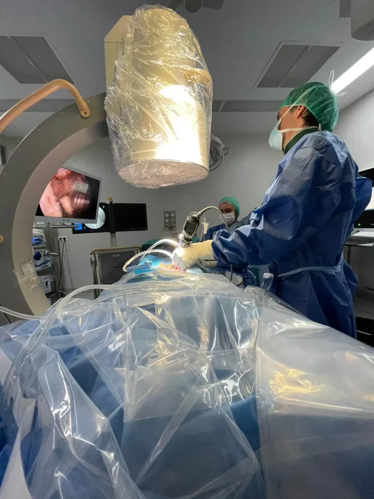
A foraminotomy aims to widen the foramen and free the root. It can be performed with open, microsurgical or endoscopic techniques in selected cases. If the narrowing is due to disc collapse, instability or spondylolisthesis, a fusion may be necessary to restore height and stabilize. In other cases, indirect decompression through interbody approaches can improve the nerve space by recovering disc height. There is no universally best technique: the best one fits the level, the cause of the narrowing, stability and the patient’s overall condition.
Benefits, risks and adverse effects
The intended benefit is to reduce irritation of the nerve root, improve radiating pain and recover the ability to walk, sleep and perform basic activities. When there is loss of strength, the goal is to halt deterioration and promote recovery, although this is not always complete.
Non-surgical treatments also have limits. Medication can cause digestive, renal or cardiovascular effects, drowsiness or dizziness. Injections can cause local pain, transient blood glucose elevation, bleeding, infection, headache or temporary nerve irritation, although serious complications are uncommon when performed with proper technique.
Surgery can involve infection, bleeding, nerve injury, cerebrospinal fluid leak, thrombosis, persistent pain, restenosis, instability or the need for reoperation. In fusions there is also a risk of nonunion, implant failure or overload of adjacent levels. These risks do not mean surgery should always be avoided, but that the indication must be careful and proportional to the problem.
Referral criteria
Specialized assessment is advisable if leg pain persists for more than 6 to 8 weeks despite a correct conservative plan, if it significantly limits walking or sleep, if there is weakness, if MRI shows severe foraminal stenosis compatible with the symptoms or if there are multiple possible diagnoses and it is unclear which nerve is causing the problem.
It is also reasonable to refer sooner when the patient is elderly, has osteoporosis, prior surgery, lumbar deformity, spondylolisthesis, rapidly progressive pain or requires strong medication to function.
Realistic recovery times
With conservative treatment, progress is usually measured in weeks. The initial goal is to reduce pain enough to walk better and regain activity. After an injection, some people notice relief within days, others within one to two weeks, and some do not respond.
After a limited or endoscopic decompression, mobilization usually begins early, but the nerve can take weeks or months to calm down. Leg pain typically improves before tingling or strength. After a fusion, timelines are longer: the first weeks focus on pain control and walking; autonomy usually increases between 6 and 12 weeks; consolidation and functional recovery can continue for several months. Physical jobs require more caution than sedentary work.
When to go to the emergency room
Go to the emergency room if new or progressive loss of leg strength appears, foot drop, inability to walk, numbness in the genital or anal area, loss of bladder or bowel control, fever with severe back pain, pain after major trauma or unbearable pain that does not improve with prescribed medication. These signs should not wait for a routine review.
Myths and realities
Myth: if there is foraminal stenosis on MRI, you must operate
Reality: many images show wear-and-tear without being the main cause of pain. Surgery is considered when imaging, symptoms and examination coincide.
Myth: all sciatica is a herniated disc
Reality: sciatica can be due to several causes. In adults with osteoarthritis or loss of disc height, foraminal stenosis is a possibility.
Myth: an injection cures the stenosis
Reality: it can reduce inflammation and pain, but it does not eliminate the structural narrowing.
Myth: a minimally invasive technique is always better
Reality: it can be very useful in selected patients, but it does not replace a good indication nor is it suitable for all anatomies.
Frequently asked questions
Is lumbar foraminal stenosis serious?
It can be mild and manageable, or very limiting if it compresses a nerve root. Severity depends on symptoms, strength, limitation to walk and correlation with imaging.
Can it be cured without surgery?
Some people improve with exercise, activity changes, cautious medication and injections. If there is persistent severe compression or progressive weakness, surgery may be considered.
Is it the same as central canal stenosis?
Not exactly. Central canal stenosis affects the central space. Foraminal stenosis affects the lateral tunnel where a root exits. They can coexist.
Does MRI always detect the problem?
MRI helps a lot, but some stenoses vary with posture or are difficult to interpret. That is why the examination and sometimes other tests matter.
What surgery is most commonly used?
It depends on the cause. It can be foraminotomy, endoscopic decompression, open decompression or fusion if there is collapse, deformity or instability.
Does strength return after freeing the nerve?
Sometimes it improves, but not always completely. Duration of compression, severity of the deficit, age, overall health and prior nerve damage influence recovery.
Can I exercise if I have foraminal stenosis?
Maintaining adapted activity is usually recommended. Exercises that trigger radicular pain or increase weakness should be avoided. A physiotherapist can tailor the plan.
When should I ask for a second opinion?
When complex surgery is proposed, there are multiple degenerated levels, symptoms do not match the MRI or you do not understand the treatment objective.
Glossary of medical terms
- Foramen: tunnel between vertebrae through which a nerve root exits.
- Stenosis: narrowing of an anatomic space.
- Radiculopathy: symptoms from irritation or compression of a nerve root.
- Sciatica: pain that travels through the buttock and leg; it is a symptom, not a single cause.
- Foraminotomy: surgery to enlarge the foramen and free the nerve.
- Spondylolisthesis: displacement of one vertebra over another.
- Decompression: procedure intended to remove pressure from nerves.
- Fusion or arthrodesis: surgery that stabilizes a segment by joining vertebrae.
References
- NICE. Low back pain and sciatica in over 16s: assessment and management. https://www.nice.org.uk/guidance/ng59. 2016, updated 2020.
- North American Spine Society. Diagnosis and Treatment of Degenerative Lumbar Spinal Stenosis. https://www.spine.org/documents/researchclinicalcare/guidelines/lumbarstenosis.pdf. 2011.
- NCBI Bookshelf. Lumbar Spinal Stenosis. https://www.ncbi.nlm.nih.gov/books/NBK531493/. 2024.
- AANS. Lumbar Spinal Stenosis. https://www.aans.org/patients/conditions-treatments/lumbar-spinal-stenosis/. 2025.
- Katz JN et al. Diagnosis and Management of Lumbar Spinal Stenosis. https://pubmed.ncbi.nlm.nih.gov/35503342/. 2022.
- Nurmukhametov R et al. Exploring Pathways for Pain Relief in Treatment and Management of Lumbar Foraminal Stenosis. https://pmc.ncbi.nlm.nih.gov/articles/PMC11352478/. 2024.
- Soar H et al. Lumbar radicular pain. https://pmc.ncbi.nlm.nih.gov/articles/PMC9402780/. 2022.
- Zaina F et al. Surgical versus non-surgical treatment for lumbar spinal stenosis. https://www.cochrane.org/evidence/CD010264_surgical-versus-non-surgical-treatment-lumbar-spinal-stenosis. 2016.
- Massachusetts General Brigham. Rehabilitation Guidelines for Conservative Management of Spinal Stenosis of the Lumbar Spine. https://www.massgeneral.org/assets/MGH/pdf/orthopaedics/sports-medicine/physical-therapy/rehabilitation-protocol-conservative-management-of-lumbar-spinal-stenosis.pdf. 2025.
Health education notice
This content is informative and does not replace an individual medical assessment. It does not allow confirmation of diagnoses or decisions about personalized treatments. If there is loss of strength, sphincter disturbance, fever, trauma or rapid worsening, seek urgent medical attention.