Thoracolumbar fusion is an operation to stabilize the spine in selected cases of advanced degeneration, spondylolisthesis, deformity, or fractures. It isn’t for everyone: it requires a thorough clinical and imaging assessment, exhausting conservative care when appropriate, and aligning expectations with a structured rehabilitation plan. Recovery is usually counted in weeks to months depending on complexity, and risks exist, although they can be reduced by Enhanced Recovery After Surgery (ERAS) protocols, navigation/robotics, and an experienced team.
- Indicated when there is instability, significant deformity, or refractory pain with imaging–clinical correlation.
- There are conservative alternatives and other surgeries (e.g., decompression, disc arthroplasty) depending on the case.
- Length of stay and complications can be reduced with ERAS and precise planning.
- Return to desk work is usually between 4–8 weeks; manual/physical jobs, 8–12+ weeks, with individual variability.
What is thoracolumbar fusion and who may benefit?
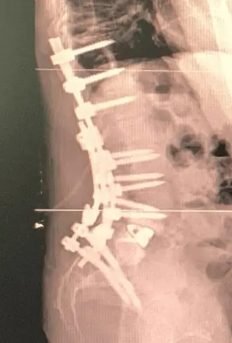
It is a procedure that permanently joins two or more vertebrae in the thoracic and lumbar regions using bone graft and implants (pedicle screws, rods and, at times, interbody cages). Its goal is to eliminate painful or unstable motion, correct alignment, and protect neural structures.
Common indications:
- Degenerative or isthmic spondylolisthesis with documented instability and disabling symptoms.
- Spinal canal stenosis with associated instability or deformity that requires stabilization in addition to decompression.
- Adult spinal deformity (scoliosis/kyphosis) that impairs function and quality of life.
- Unstable fractures, tumor, or infections that compromise stability.
- Pseudoarthrosis (nonunion after a prior fusion) or previous surgery with mechanical failure.
Clinical guidelines and coverage criteria from scientific societies stress careful selection: fusion should not be offered for nonspecific low back pain without instability or deformity, nor as an automatic response to an isolated disc herniation. The key is correlating symptoms and imaging and exhausting conservative options when indicated.
How is it diagnosed and planned?
The decision relies on:
- History and neurological examination: pain pattern, neurogenic claudication, motor or sensory deficits, functional impact.
- MRI: assesses stenosis, disc disease, and neural compression.
- Dynamic X-rays (flexion–extension): reveal instability in some cases.
- CT: useful to evaluate facets, pars interarticularis, prior fusion, or complex osteotomies.
- 3D planning and navigation/robotics: improve implant accuracy and may reduce complications.
In specific scenarios (pure discogenic pain, facet pain, sacroiliac pain), additional tests (diagnostic blocks) may be considered if they will genuinely change management.
Alternatives: conservative and surgical
Before surgery
- Therapeutic exercise and education (biopsychosocial approach): the foundation of low back pain care and many compensated deformities.
- Judicious medication (non-opioid analgesics and short courses of anti-inflammatories; avoid chronic opioids).
- Active physiotherapy; passive modalities as adjuncts.
- Injections/radiofrequency ablation: in selected cases (facet or sacroiliac).
- Regenerative medicine in restricted indications (PRP/mesenchymal cells) when aiming to delay or avoid surgery; evidence is heterogeneous and must be individualized.
Surgical options other than thoracolumbar fusion
- Isolated decompression (laminectomy/foraminotomy) when there is stenosis without instability.
- Disc arthroplasty (highly selected cases, usually one or two levels and without relevant instability) when preserving motion is the goal.
- Minimally invasive approaches (MIS) —OLIF/XLIF, TLIF/PLIF MIS— which can be integrated into fusion strategies with less soft-tissue disruption.
The choice combines diagnosis, anatomy, functional goals, and patient expectations.
Expected benefits versus risks and adverse effects
Potential benefits: relief of pain of mechanical/instability origin, improved walking and functional capacity, deformity correction, neurological protection, and—when well indicated—durable outcomes.
Risks and complications: infection, bleeding/thrombosis, cerebrospinal fluid leak, nerve/vascular injury, malposition or breakage of implants, nonunion, persistent pain, anesthetic complications. The likelihood depends on age, comorbidity, smoking, bone density, number of levels fused, and the complexity of correction.
How to reduce risks: ERAS protocols, preoperative optimization (anemia, nutrition, glycemic control, smoking cessation), 3D planning, navigation/robotics, and early rehabilitation. Recent literature associates ERAS with shorter stays, less postoperative pain and, in some studies, fewer complications.
Realistic recovery timelines
- First 48–72 h: multimodal pain control, early mobilization, discharge when pain, walking, and home safety allow.
- Weeks 2–4: daily walks, posture hygiene; start of structured physiotherapy (depending on surgery and progress).
- Weeks 4–8: gradual return to desk work if pain is controlled and without sedating medication; core strengthening.
- Weeks 8–12: increase endurance and strength; many patients with physical jobs need a gradual ramp-up and additional weeks.
- 3–6 months: functional consolidation; higher-impact activities only if the surgeon authorizes them based on fusion and clinical status.
These timeframes vary with the number of levels, need for osteotomies, bone quality, age, and fitness. Expectations should be adjusted in clinic.
Practical criteria for referral for surgical evaluation
- Disabling pain or neurogenic claudication with work/family impact despite ≥6–12 weeks of well-delivered treatment.
- Progressive neurological deficit or significant radiological instability.
- Deformity that alters sagittal/coronal balance and quality of life.
- Failed fusion (pseudoarthrosis) with mechanical pain and/or dysfunction.
When should I go to the emergency department?
- Progressive leg weakness, foot drop, or sudden loss of strength.
- High fever with severe back pain after surgery.
- Changes in bladder/bowel control or saddle anesthesia.
- Sudden, incapacitating pain with rapid worsening.
Myths and facts
- Myth: “Fusion cures any low back pain.” Fact: it’s only useful for specific indications; not for nonspecific pain without instability or deformity.
- Myth: “If I have surgery, I can forget about physiotherapy.” Fact: exercise and rehabilitation are key determinants of the outcome.
- Myth: “MIS techniques eliminate risk.” Fact: they can reduce tissue trauma but don’t remove inherent risks.
FAQs
In which cases is fusion usually not recommended?
Nonspecific low back pain without instability, stenosis without clear instability, or an isolated disc herniation without fusion criteria are usually managed with other options.
How long does surgery take and how many levels are typically fused?
From 2 to 5 hours depending on levels and complexity. The number of levels depends on the condition; deformity may require multiple levels, spondylolisthesis sometimes 1–2.
Do navigation or robots guarantee better results?
They help place implants more accurately and standardize processes, which may reduce complications. They don’t replace appropriate case selection or surgical experience.
When can I drive and return to work?
Short drives when pain allows and without sedating medication (usually from 3–4 weeks). Desk work 4–8 weeks; physical work, 8–12+ depending on recovery.
Is mobility lost forever?
Movement at fused levels is sacrificed, but the aim is to preserve overall function and relieve pain. Rehabilitation helps compensate.
What’s the chance of reoperation?
There’s a risk of nonunion or adjacent-segment problems in the mid- to long-term. Proper selection, sound technique, and healthy habits reduce that risk.
Glossary
- Arthrodesis: surgical fusion of two or more vertebrae.
- Osteotomy: controlled bone cut to correct a deformity.
- ERAS: enhanced recovery protocol to optimize the postoperative course.
- Pseudoarthrosis: lack of bony consolidation after a fusion.
- MIS approach: minimally invasive technique with less tissue disruption.
Notice: This content is informational and does not replace an individual medical evaluation.
References
- North American Spine Society (NASS). Coverage & Clinical Guidelines on lumbar fusion and use criteria (updates 2021–2025). https://www.spine.org (accessed 2025).
- Congress of Neurological Surgeons (CNS). Lumbar Fusion Guideline (J Neurosurg Spine). 2014, with updates and complementary literature. https://www.cns.org (accessed 2025).
- NICE NG59. Low back pain and sciatica in over 16s: assessment and management. https://www.nice.org.uk/guidance/ng59 (accessed 2025).
- Debono B, et al. Consensus statement for perioperative care in lumbar spinal surgery (ERAS). Spine J. 2021. https://www.thespinejournalonline.com (accessed 2025).
- Bansal T, et al. ERAS in spine surgery: review. 2022. https://pmc.ncbi.nlm.nih.gov/articles/PMC9293758/ (accessed 2025).
- Xu W, et al. Meta-analysis: lumbar fusion vs non-surgical management in chronic pain. 2021. https://pubmed.ncbi.nlm.nih.gov/33253955/ (accessed 2025).
Disclaimer: This content is educational and does not replace an individual medical evaluation. If you have concerning symptoms or questions about your case, consult a healthcare professional.