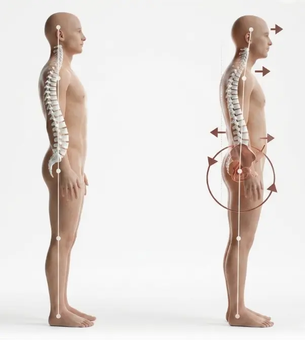
Flat back syndrome occurs when the lumbar region loses part of its natural curve and the body leans forward. It can result from degeneration, fractures or after a spinal fusion. Not every stooped posture requires surgery, but pain, fatigue and loss of independence justify a full assessment of alignment.
- The most characteristic sign is difficulty staying upright without flexing the hips or knees.
- Diagnosis combines symptoms, examination and standing full-spine X-rays.
- Physiotherapy and pain control can improve function, although they do not straighten a rigid deformity.
- Surgery is reserved for selected cases with significant disability and a reasonable balance between benefits and risks.
- New weakness, loss of sphincter control or fever with pain require urgent attention.
What is flat back syndrome
Seen from the side, a healthy spine is not straight. The lumbar region has an inward curve called lumbar lordosis. This curve positions the head over the pelvis and allows standing with reasonable muscle effort. In flat back syndrome lordosis is lost and the trunk shifts forward. It is also called sagittal imbalance when the alteration affects the body’s alignment in the lateral plane.
To keep the gaze horizontal, the body compensates by extending the neck, moving the pelvis backward and flexing hips and knees. These strategies can hide the deformity in a photograph but require continuous effort. That is why some people appear relatively straight at the start of the day and end up increasingly bent and tired.
It may appear after a lumbar fusion performed with insufficient lordosis, due to degeneration of discs and muscles, vertebral fractures, osteoporosis, Parkinson’s disease or other neuromuscular disorders. It can also coexist with scoliosis, stenosis, pseudoarthrosis or deterioration of adjacent segments. Having a visually straight back on an X-ray is not enough to diagnose the syndrome: there must be a measurable alteration and related symptoms.
Symptoms and indications: 9 useful signs
The following signs do not confirm the diagnosis by themselves but help recognize a pattern that warrants evaluation:
- Progressive forward leaning. It becomes difficult to keep the shoulders and head over the pelvis, especially when standing.
- Need to bend the knees. Flexion allows looking forward but overloads the thighs and joints.
- Fatigue when walking. Tolerated distance decreases and there may be a need to lean on a trolley, cane or countertop.
- Mechanical low back pain. It usually increases with standing or at the end of the day and improves when sitting or lying down.
- Pain in the thighs or knees. It does not always originate in those joints; it can reflect the constant compensatory effort.
- Difficulty looking forward. Maintaining a horizontal gaze forces neck extension and can cause neck pain.
- Loss of height or postural change. Clothes fit differently and family members may notice that the trunk has shifted forward.
- Limitation of daily activities. Cooking, standing in line, showering or walking become exhausting tasks.
- Associated nerve symptoms. Leg pain, tingling, loss of strength or heaviness may indicate stenosis or added compression.
Not every stoop is flat back. A flexible posture improves when lying down or with voluntary correction. A structural deformity can persist even at rest. In addition, hip or knee disease, Parkinson’s or muscle weakness can mimic it. Distinguishing these causes changes management.
How it is diagnosed without relying on a single image
Evaluation begins with the medical history: previous surgeries, evolution of posture, pain, walking distance, falls, smoking, medications, osteoporosis and neurological diseases. It is useful to describe what happens at the start and at the end of the day. Examination observes gait, balance, pelvis and knee position, curve flexibility, strength, sensation and reflexes.
The basic test is usually a standing full-spine X-ray that includes the head, pelvis and, when possible, the femurs. It allows measurement of spinopelvic alignment, that is, the relationship between the spine and pelvis. Parameters used include the sagittal vertical axis, pelvic tilt and the difference between pelvic incidence and lumbar lordosis. There is no single ideal number for all ages: overcorrection can also be harmful.
Flexion and extension radiographs help determine whether the deformity is flexible. CT shows fusion consolidation, screws, fractures and the structural quality of the bone. MRI allows assessment of discs, nerves, stenosis, infection or tumor. If reconstruction is considered, bone density by DXA or CT, vitamin D, nutrition, cardiopulmonary function and frailty may need to be evaluated.
A radiological finding is only relevant when it explains the symptoms and the examination. The goal is not to pursue a perfect measurement but to determine what part of the disability comes from the imbalance and which coexisting problems also need treatment.
Non-surgical and surgical options
Non-surgical treatment
In mild, flexible deformities or when surgical risk is high, conservative treatment can improve endurance, pain and independence. It usually includes individualized physiotherapy to strengthen trunk extensors, glutes and legs, work on balance and maintain hip mobility. Exercise should be adapted to osteoporosis, stenosis and neurological symptoms.
Postural education, scheduled breaks, walking aids and task adaptation may also be used. Analgesics are considered according to age, kidney, stomach, cardiovascular risk and other medications. Injections can treat a specific source of radicular or facet pain but do not correct a rigid imbalance. A brace may provide temporary support in selected cases, although prolonged use can promote weakness and does not by itself restore lost lordosis.
It is important to treat osteoporosis, vitamin D deficiency, malnutrition, smoking and muscle mass loss. These measures help even if surgery is never performed and reduce risks if an intervention is eventually considered.
Surgical options
Surgery is considered when the deformity causes severe limitation, progresses or is accompanied by neurological compression, and conservative treatment does not provide acceptable function. Planning is personalized. It may include removing or revising implants, decompressing nerves, revising a nonunited fusion, placing interbody cages to restore height and lordosis or extending instrumentation.
A flexible deformity can be corrected using interbody approaches and posterior osteotomies. An osteotomy is a controlled bone cut to change alignment. Rigid deformities may require a pedicle subtraction osteotomy, more powerful but also more complex. Not everyone needs the same correction or a fusion down to the pelvis.
Benefits, risks and adverse effects
The intended benefit is to recover a more sustainable posture, reduce pain and fatigue, increase walking distance and facilitate basic activities. A nerve can also be decompressed or an unstable area stabilized. Improvement does not always mean complete pain relief and can take months.
Reconstruction of an adult deformity is major surgery. Risks include bleeding and transfusion, infection, thrombosis, pulmonary or cardiac problems, nerve injury, cerebrospinal fluid leak, loss of strength, persistent pain and anesthetic complications. In the medium term there may be nonunion, loosening or breakage of implants, cage subsidence, fracture, proximal junctional kyphosis, loss of correction and repeat surgery.
Individual risk changes with biological age, frailty, obesity, diabetes, smoking, osteoporosis, nutrition, number of prior surgeries and extent of reconstruction. Therefore the decision must compare the current impact of the deformity with a realistic scenario of benefit, complications and recovery.
Criteria for specialist referral
Consider a scheduled specialist assessment if the forward lean progresses, walking or standing becomes increasingly difficult, knee and hip compensations appear, there is persistent pain after a fusion or X-rays show significant loss of lordosis. Also if well-directed conservative treatment does not allow maintaining essential activities.
Referral should be prioritized in the presence of loss of strength, worsening gait disturbance, repeated falls, unexplained night pain, rapid deterioration or suspected implant failure. Bringing surgical reports and prior studies allows comparison of alignment over time.
Realistic recovery times
With conservative treatment, functional changes are usually assessed after several weeks or months of consistent work. Tolerance to effort can improve even if the X-ray changes little.
After reconstruction, getting up and walking with assistance usually begins during the first days if the situation allows. Hospital stay may last several days and some people need additional rehabilitation. The first six weeks focus on pain control, wound care, progressive walking and avoiding loads or movements not authorized.
Between six and twelve weeks independence increases, but fatigue remains common. Sedentary work may require six to twelve weeks or more. Physical jobs often demand several months. Bone consolidation and muscle recovery continue for six to twelve months, sometimes longer after extensive revisions. These figures are indicative: a complication, frailty or a very long surgery can lengthen each phase.
When to go to the emergency department
Chronic forward posture is rarely an emergency by itself. Go to the emergency department if rapid weakness appears in one or both legs, new inability to walk, loss of bladder or bowel control, urinary retention or numbness in the genitals and perineum. Also seek urgent care for fever with intense back pain or a surgical wound, sudden pain after a fall, a deformity that changes suddenly or pain with a history of cancer and rapid deterioration.
Myths and realities
- Myth: it is just bad posture. Reality: there can be a structural loss of lordosis that is not voluntarily corrected.
- Myth: every flat back needs surgery. Reality: symptoms and disability are treated, not an isolated X-ray.
- Myth: strengthening will always straighten the spine. Reality: it improves capacity and control but does not remodel a rigid fusion.
- Myth: surgery guarantees a perfect posture. Reality: the aim is a proportionate, functional alignment with acceptable risks.
- Myth: more correction is always better. Reality: the goal must consider age, anatomy and adaptive capacity.
Frequently asked questions
Does flat back always appear after surgery?
No. It can also be due to degeneration, fractures, osteoporosis or neuromuscular disorders. Prior surgery, especially a long fusion, is a known cause but not the only one.
How do I know if it is bad posture or a fixed deformity?
A flexible posture usually corrects when lying down or with conscious effort. A fixed deformity persists and produces compensations. Examination and standing and dynamic X-rays help differentiate them.
Does an MRI diagnose sagittal imbalance?
It is not usually the main test because it is typically performed lying down. Weight-bearing full-spine radiographs show global alignment better. MRI provides information about nerves, discs and stenosis.
Can physiotherapy prevent surgery?
It can improve strength, balance, pain and exercise tolerance, and in some people allow avoiding or postponing surgery. It usually does not correct a poorly aligned rigid fusion.
When is revision surgery considered?
When there is significant disability, progression, mechanical pain or neurological compression that fit the deformity, after weighing alternatives, risks and general condition. It is not indicated solely for a radiographic measurement.
Is there an upper age limit for surgery?
There is no universal number. Frailty, cardiopulmonary health, bone quality, nutrition, independence and the magnitude of surgery matter more than chronological age alone.
Will I walk upright immediately?
Alignment changes during surgery, but walking naturally requires regaining strength, balance and confidence. Fatigue and walking aids are common at first.
Can it recur?
Part of the correction can be lost or a problem can arise in adjacent areas, especially with fragile bone or extensive fusions. Planning, medical optimization and follow-up reduce risks but do not eliminate them.
Glossary of medical terms
- Lumbar lordosis: natural inward curve of the lower back.
- Sagittal balance: the body’s alignment observed from the side.
- Pelvic incidence: an anatomical measure of the pelvis used to plan alignment.
- Fusion or arthrodesis: surgical joining of two or more vertebrae.
- Pseudoarthrosis: failure of a fusion to consolidate.
- Osteotomy: a planned bone cut to correct a deformity.
- Stenosis: narrowing of the space available for nerves or the spinal cord.
- Proximal junctional kyphosis: abnormal curvature near the upper end of a fusion.
References
- Scoliosis Research Society. Post-Surgical Malalignment. https://www.srs.org/Patients/Conditions/Post-Surgical-Malalignment Accessed 2026.
- Scoliosis Research Society. Surgery: Fixed Sagittal Imbalance. https://www.srs.org/Patients/Diagnosis-And-Treatment/Surgery Accessed 2026.
- Hospital for Special Surgery. Revision Surgery for Spine Deformity in Adults. https://www.hss.edu/health-library/conditions-and-treatments/scoliosis-kyphosis-revision-surgery-spinal-deformity Year 2023.
- Kim HJ and colleagues. Adult Spinal Deformity: A Comprehensive Review. https://pubmed.ncbi.nlm.nih.gov/36274246/ Year 2022.
- Kim HJ and colleagues. Adult Spinal Deformity: Current Concepts and Decision-Making Strategies. https://pmc.ncbi.nlm.nih.gov/articles/PMC7788366/ Year 2020.
- Cheung JPY. The importance of sagittal balance in adult scoliosis surgery. https://pmc.ncbi.nlm.nih.gov/articles/PMC6995914/ Year 2020.
- Taşkıran ÖÖ. Rehabilitation in adult spinal deformity. https://pubmed.ncbi.nlm.nih.gov/33089079/ Year 2020.
- Glassman SD and colleagues. The impact of positive sagittal balance in adult spinal deformity. https://pubmed.ncbi.nlm.nih.gov/16166889/ Year 2005.
- Teles AR and colleagues. Effectiveness of Operative and Nonoperative Care for Adult Spinal Deformity. https://pubmed.ncbi.nlm.nih.gov/28507887/ Year 2017.
- Congress of Neurological Surgeons. Preoperative Osteoporosis Assessment. https://www.cns.org/guidelines/browse-guidelines-detail/3-preoperative-osteoporosis-assessment Year 2021.
- NICE. Low back pain and sciatica in over 16s: assessment and management, NG59. https://www.nice.org.uk/guidance/ng59 Year 2016, updated 2020.
- Kim JS and colleagues. Surgical Outcomes of Post-Fusion Lumbar Flatback Deformity. https://pmc.ncbi.nlm.nih.gov/articles/PMC5106362/ Year 2016.
Health education notice
This content is educational and does not replace an individual medical assessment or allow a diagnosis to be established. Treatments depend on symptoms, examination, imaging and overall condition. In the presence of alarm signs or rapid deterioration, urgent medical attention should be sought.