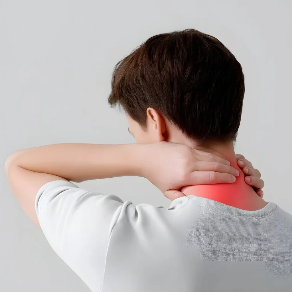
Cervicothoracic deformity is a misalignment between the neck and the upper back that can cause pain, difficulty keeping the gaze level, and—in advanced cases—spinal cord compression. Management requires meticulous clinical and imaging assessment: defining the deformity type and its stiffness, and weighing when conservative care is enough and when surgical correction (sometimes using osteotomies) brings more benefit than risk. This article, written in clear language and based on recent guidelines and reviews, explains symptoms, useful tests, alternatives, benefits and complications, referral criteria, and realistic recovery timelines.
- Not every deformity needs surgery: the goal is to restore function and a safe forward gaze.
- Decisions depend on deformity type, stiffness, and neurological/functional impact.
- Surgery may require osteotomies at the cervicothoracic junction; it offers improvements but carries specific risks.
- Recovery is measured in months; the rehabilitation plan and bone strength make the difference.
What is cervicothoracic deformity?
It is a disturbance of the balance between the cervical and thoracic spine at the cervicothoracic junction (C7–T1). It may present as a forward-tilted head, inability to maintain a horizontal gaze (increased chin–sternum angle or “chin–brow vertical angle”), upper-neck/upper-back pain and, at times, tingling or weakness due to spinal cord or nerve-root compromise. It is often associated with disc degeneration, ankylosing spondylitis, postoperative sequelae (e.g., kyphosis after laminectomy), scoliosis, or neuromuscular disorders.
Symptoms and when to seek evaluation
- Cervical/upper thoracic pain that worsens when keeping the head upright or looking straight ahead.
- Difficulty maintaining a horizontal gaze or swallowing due to forced postures.
- Tingling, weakness or hand clumsiness; gait disturbance.
- Marked functional limitation in activities of daily living.
When to prioritize work-up: rapid symptom progression, dropping objects due to weakness, sphincter dysfunction, or frequent falls.
Diagnosis: tests that actually help
History and neurological examination guide the process. Full-length standing radiographs (with flexion/extension views) are key to measure parameters such as cervical lordosis, T1 slope, and the chin–brow vertical angle (CBVA). MRI evaluates spinal cord/foraminal compression, and CT assesses deformity stiffness and bone quality. In selected cases, dynamic studies or standardized functional scales are added.
Treatment options
Non-surgical
- Education and targeted physiotherapy (posture, scapular/cervical mobility, progressive strengthening).
- Pain control (rational analgesia; avoid long-term opioids unless specifically indicated).
- Management of comorbidities (osteoporosis, sarcopenia) and nutritional optimization.
- Targeted injections in cases with predominantly facet or radicular pain without severe neurological compromise.
Surgical
Considered when there is a neurological deficit, a rigid deformity with major functional impact, refractory pain, or inability to maintain a forward gaze despite appropriate conservative care. Techniques include:
- Decompression (anterior and/or posterior) when there is cord/foraminal compression.
- Fixation and instrumentation to restore and maintain corrected alignment.
- Osteotomies (controlled opening/closing cuts through vertebrae) in rigid deformities; at the cervicothoracic junction, three-column osteotomies may be required to gain sagittal correction.
Benefits vs. risks (what we know in 2025)
Expected benefits
- Pain reduction and improved ability to look straight ahead and hold a functional posture.
- Decrease in neurological symptoms when compression was present.
- Improvements in quality of life and independence in daily activities.
Risks and adverse effects
- Respiratory complications, dysphagia/dysphonia (cervical approaches), infection, or cerebrospinal fluid (CSF) leak.
- Neurological injury (including C5 palsy), implant failure, or partial loss of correction.
- Need for reoperation in a proportion of cases—higher with poor bone quality, malnutrition, or extensive deformities.
Actual complication risk varies with age, comorbidities, osteotomy type, and construct length. Optimizing bone health, muscle strength, and anemia pre-op reduces risk.
Practical criteria for referral to a spine unit
- Inability to maintain a horizontal gaze that compromises safety when walking or driving.
- Signs of myelopathy (hand clumsiness, falls, sphincter changes) or progressive radiculopathy.
- Failure of well-structured conservative care over 8–12 weeks with high functional impact.
- Rigid deformity at the cervicothoracic junction with disabling pain.
Realistic recovery timelines
- Hospital: 3–7 days depending on surgery magnitude and comorbidities.
- First month: early ambulation, collar if indicated, pain control, gentle exercises.
- 6–12 weeks: ramp-up physiotherapy (scapular mobility, endurance), progressive return to sedentary work.
- 3–6 months: functional consolidation; gradual return to physically demanding activities.
- >6 months: assess bony fusion and stability of the correction.
When to seek urgent care?
- Progressive weakness, widespread sensory loss, or sphincter dysfunction.
- High fever with severe pain, or a surgical wound that is red and draining.
- New chest pain or breathing difficulty.
Myths & facts
- Myth: “Surgery makes everything perfectly straight forever.” Fact: correction seeks function and safety; it may require long constructs and later adjustments.
- Myth: “If I have surgery, I won’t be able to move my neck.” Fact: motion depends on the level and type of fusion; non-fused segments retain movement and compensatory training is used.
- Myth: “Osteotomies are always extremely dangerous.” Fact: they carry specific risks, minimized with planning, neuromonitoring, and pre-operative optimization.
FAQs
Is surgery always necessary for a cervicothoracic deformity?
No. Many people improve with structured physiotherapy and pain control. Surgery is considered when there is neurological deficit, a rigid deformity with major functional impact, or failure of conservative management.
Which tests are essential before deciding?
Full-length standing radiographs (with flexion/extension) to measure alignment, MRI to assess neurological compression, and CT to estimate stiffness and plan osteotomies if needed.
What is a three-column osteotomy?
A powerful correction that opens/closes the bone through the entire vertebra to restore alignment. It is reserved for rigid deformities, especially at the cervicothoracic junction.
How soon can I return to work?
For sedentary jobs, often between 6 and 12 weeks; with physical demands, 3–6 months may be needed, depending on progress, fusion, and surgery type.
Does osteoporosis preclude surgery?
Not always, but bone health must be optimized and fixation strategy adapted to reduce loosening or implant failure.
Does correction “cure” pain 100%?
Total elimination of pain cannot be guaranteed. The goal is to improve function, safety, and quality of life; residual pain depends on many factors.
Glossary
- CBVA: chin–brow vertical angle reflecting the ability to keep a horizontal gaze.
- T1 slope: parameter relating T1 orientation to the cervical lordosis required.
- Osteotomy: controlled bone cut to correct a deformity.
- Cervicothoracic junction: transition between cervical and thoracic spine (C7–T1).
References
- Cervicothoracic deformity correction: everything you need to know. Dr. Vicenç Gilete, MD, Neurosurgeon, and Dr. Augusto Covaro, MD, Orthopedic Surgeon. Complex Spine Institute, Barcelona, Apr 18, 2025:
https://complexspineinstitute.com/en/treatments/cervicothoracic-deformity-correction/ - Passias PG, et al. Current concepts in adult cervical spine deformity surgery (J Neurosurg Spine, 2024). https://thejns.org/spine/view/journals/j-neurosurg-spine/40/4/article-p439.xml
- Kaidi AC, et al. Classification(s) of Cervical Deformity (Neurospine/PMC, 2022). https://pmc.ncbi.nlm.nih.gov/articles/PMC9816582/
- Lobão A, et al. Osteotomies for the Correction of Cervical Deformity: When, How, and Why (J Spinal Disord Tech, 2025). https://journals.lww.com/jspinaldisorders/fulltext/2025/11000/osteotomies_for_the_correction_of_cervical.6.aspx
- Martínez FA, et al. Updates on the prevention and treatment of cervicothoracic deformity (Asian Spine Journal, 2025). https://asj.amegroups.org/article/view/104458/html
- Shah I, et al. Effect of Osteoporosis on Outcomes After Surgery for Cervical Deformity (J Clin Med, 2025). https://www.mdpi.com/2077-0383/14/17/6196
- Mikula AL, et al. Neurological outcomes after three-column osteotomy: level selection in cervical deformity (J Neurosurg Spine, 2025). https://thejns.org/spine/view/journals/j-neurosurg-spine/43/4/article-p433.xml
This content is informational and does not replace individual medical evaluation.
