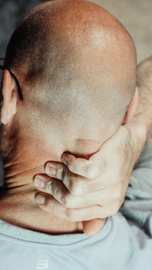
Cervical MRI is a very useful test when symptoms suggest involvement of a nerve root, the spinal cord, or a structural lesion that would change medical management. But it does not always add value: in nonspecific neck pain without red flags, it can show common findings that do not explain the problem and may confuse more than help. Knowing when to request a cervical MRI and when not to avoids unnecessary tests, falsely alarming findings, and delays in appropriate treatment.
- Cervical MRI sees soft tissues better: discs, the spinal cord, nerves and ligaments.
- It is not the first test for every case of neck pain.
- It is more useful when there is radiculopathy, myelopathy or red flags.
- X-ray assesses alignment and bone better; CT, bony detail and trauma.
- An MRI can show “age-related” findings that do not cause symptoms.
- The imaging report must always be interpreted together with the clinical examination.
- If symptoms progress or there is neurological deficit, the indication changes.
What a cervical MRI is and why there is doubt about ordering it
The cervical MRI, also called magnetic resonance imaging (MRI) of the cervical spine, is an imaging test that uses a magnetic field and radio waves to obtain detailed slices of the neck. It allows very precise evaluation of the intervertebral discs, the spinal cord, nerve roots, ligaments and other soft tissues that are not well seen on an X-ray. In addition, it does not expose the patient to ionizing radiation.
The doubt about when to request a cervical MRI and when not to arises because neck pain is common, but not all neck pain means a serious lesion. Many complaints improve with conservative measures. In addition, cervical MRI detects very common age-related changes, such as disc protrusions, osteoarthritis or small narrowings, that can be present even in people without symptoms. For that reason, an “abnormal” image does not automatically equate to a clear cause of pain.
That is the clinical key: a cervical MRI is not ordered just to “see if there is something,” but when the information it can provide changes specific decisions. It can help confirm nerve compression, assess suspected myelopathy, study trauma with neurological symptoms, or rule out infection, tumor or inflammation when the clinical history suggests it.
The confusion is worsened because the cervical spine ages like other body structures. It is normal for degenerative changes to appear over the years, but many do not produce pain. Therefore, the challenge is not only whether to order a cervical MRI, but to order it at the right time and for the right reason.
Symptoms and warning signs: when cervical MRI really adds value
In mild or moderate neck pain without neurological symptoms or relevant trauma, cervical MRI usually has limited yield at the outset. However, there are symptoms that do change the threshold for imaging:
Pain radiating down the arm
When cervical pain radiates to the shoulder, arm, forearm or hand, there may be cervical radiculopathy, that is, irritation or compression of a nerve root exiting the neck. If it is accompanied by pins and needles, numbness or loss of strength, cervical MRI is usually much more useful.
Weakness, clumsiness or loss of dexterity
Weakness in an arm, difficulty buttoning a shirt, fine hand clumsiness or a sensation of a “strange hand” should raise concern for neurological involvement. If there is suspicion of cervical myelopathy, that is, compression of the spinal cord in the neck, cervical MRI ceases to be optional and becomes a highly relevant test.
Gait or balance disturbance
A stiffer, insecure or clumsy gait, especially if accompanied by hyperreflexia, loss of balance or a sensation of “odd” legs, may indicate spinal cord compromise. In that context, cervical MRI helps identify cord compression and assess the urgency of treatment.
Trauma with neurological symptoms
After a fall, accident or whiplash injury, the choice between X-ray, CT and cervical MRI depends on the scenario. If there is persistent pain, neurological deficit or suspicion of ligamentous or spinal cord injury, MRI gains weight. CT is usually better for fractures; MRI, for soft tissues and the cord.
Fever, unexplained weight loss or persistent nocturnal pain
Fever, unexplained weight loss, pain that does not improve at night, or a history of cancer require ruling out infection or tumor. In those cases, cervical MRI can be the most informative test because it visualizes the spinal cord, canal and soft tissues in great detail.
Sphincter disturbances or advanced spinal cord symptoms
Although not common in cervical pathology, loss of sphincter control, very unstable gait or progressive weakness should be treated as warning signs. If the clinical picture suggests spinal cord compression, cervical MRI should be expedited.
How it is diagnosed: physical exam, X-ray, CT and cervical MRI
Correct diagnosis does not start with imaging, but with history and neurological examination. The doctor asks when the pain began, whether there was trauma, whether the pain radiates to the arm, and whether there is tingling, loss of strength, hand clumsiness or gait changes. Then they examine neck mobility, sensation, strength, reflexes and signs of spinal cord or root compression.
The cervical X-ray is useful to see alignment, curvatures, obvious instability, marked osteoarthritis, indirect disc space narrowing and some fractures. It is a simple and inexpensive test, but it does not show discs, the spinal cord or nerve roots well. Therefore, in many cases of neck pain without red flags, it can be a reasonable first step, although it does not resolve the entire clinical question.
The cervical CT provides very good bony detail and is especially useful in trauma, fractures, bony deformities or when the X-ray does not clarify a structural suspicion. However, CT is inferior to cervical MRI for assessing the cord, nerves, discs and ligaments. In addition, it uses ionizing radiation.
The cervical MRI is the best test to study discs, roots, the vertebral canal, the spinal cord and soft tissues. It is the preferred option when radiculopathy with neurological deficit, myelopathy, infection, tumor, inflammation or soft tissue injury is suspected. But its high sensitivity also explains part of the problem: it detects many incidental findings, that is, alterations that are not necessarily the cause of symptoms.
Therefore, the most important criterion is not whether the cervical MRI “looks bad,” but whether the finding fits the patient’s side, level and clinical pattern. A protrusion on MRI may mean nothing if there is no correlation with physical examination.
Non-surgical treatment alternatives
When there are no warning signs, many causes of neck pain improve without surgery and often without an immediate cervical MRI. Conservative treatment usually includes postural education, temporary activity modification, analgesics or anti-inflammatories if indicated, local heat, therapeutic exercise and guided physiotherapy.
If there is suspected cervical radiculopathy without severe deficit, clinical observation and initial conservative treatment may be considered. During that period, cervical MRI is reserved for lack of improvement, the appearance of neurological signs, or when the result will change the therapeutic strategy.
In some patients, injections or selective nerve blocks may be part of management, especially when trying to confirm a symptomatic level or relieve resistant pain. In any case, the indication is usually supported by good clinical and imaging correlation, not by an isolated cervical MRI.
An important point is that physiotherapy does not compete with cervical MRI. They are different tools. The first treats symptoms and function; the second clarifies anatomy when the clinical suspicion justifies it.
Surgical alternatives
Surgery does not depend only on whether the cervical MRI shows a herniation or osteoarthritis. The surgical decision arises when there is correlation between symptoms, exam and imaging findings, and when conservative treatment has not been sufficient or there is neurological risk.
Options vary depending on the problem: anterior or posterior decompression, discectomy, foraminotomy or procedures to decompress the spinal cord. In some cases fusion (arthrodesis) is added to provide stability. Cervical MRI helps define the level, side and type of compression, but it does not decide surgery on its own.
If the clinical picture is myelopathy, the priority is not to delay specialist assessment. In contrast, if the MRI finding is mild and does not explain the symptoms, surgery may not be indicated even if the image seems striking to the patient.
Benefits, risks and adverse effects
The major benefit of cervical MRI is its ability to detect soft tissue lesions and neurological compression in great detail. It also avoids radiation and provides essential information when problems are suspected that an X-ray cannot reveal.
But it also has limitations. The main risk is often interpretive rather than physical: cervical MRI can show disc protrusions, disc dehydration, osteophytes or narrowings that do not cause symptoms. If these are interpreted out of context, they can generate anxiety, overtreatment or unnecessary decisions.
Other drawbacks are claustrophobia, noise, scan duration and limitations in patients with certain non-compatible implants or devices. In some cases contrast may be required, but not always. Also, a normal cervical MRI does not completely rule out mechanical or muscular pain, because there are many painful conditions that do not leave a clear mark on imaging.
Therefore, a practical idea to remember is: cervical MRI adds more when it answers a specific clinical question than when it is ordered as a reassurance test without a defined indication.
Referral criteria to a specialist
Consider referral to a spine specialist, neurosurgeon, orthopedic surgeon or neurologist when neck pain is accompanied by neurological symptoms, when it persists despite initial treatment, or when the examination suggests root or cord compression. Also consider referral if the patient has a history of cancer, infection, significant trauma or inflammatory diseases that lower the threshold for imaging.
Cervical MRI is usually especially useful in three scenarios: first, when there is radiculopathy with radiating pain and/or deficit; second, when myelopathy is suspected; and third, when red flags are present. If the condition is stable, without deficit and without alarm signs, it may be reasonable to start conservative management before ordering it.
A practical rule is simple: if the result of the cervical MRI can change treatment, the test is more justified. If it will not modify anything, it is probably better to wait or choose another strategy.
Realistic recovery times
Recovery depends on the cause, not the image. Mechanical neck pain can improve in days or weeks with conservative measures. Radiculopathy can take longer, sometimes several weeks or months, and the course is highly variable. Myelopathy follows a different logic because the goal is not only to relieve pain but to halt neurological deterioration.
After a cervical MRI, recovery is immediate because the test is not therapeutic. What changes is the diagnostic plan: if the image confirms relevant compression, treatment may be accelerated; if it shows mild or incidental findings, conservative options may be reinforced.
In practice, many patients believe that “having an MRI” equals “having a definitive diagnosis.” This is not always the case. The final diagnosis is a synthesis of symptoms, exam and tests. Cervical MRI contributes only a part of that story.
When to go to the emergency department
There are situations in which you should not wait for a scheduled appointment or an outpatient cervical MRI. Go to the emergency department if neck pain appears after an accident with suspected serious injury, if there is progressive weakness in an arm or leg, if there is difficulty walking or marked loss of balance, or if there is loss of sphincter control.
Other reasons for urgent assessment include fever associated with severe neck pain, a history of cancer with new persistent pain, sudden loss of strength, increasing numbness or disproportionate pain that does not respond and worsens rapidly. In those cases, cervical MRI may be part of the urgent or semiurgent workup, depending on the clinical situation.
Myths and realities
Myth: “If the neck hurts a lot, you always need a cervical MRI”
Reality: pain alone does not automatically justify the test. Cervical MRI is more useful when there is radiation to the arm, neurological deficit, trauma or suspicion of a specific cause.
Myth: “If the cervical MRI looks bad, the cause is already clear”
Reality: many findings are common in people without symptoms. The key is that the image fits the clinical picture and examination.
Myth: “X-rays are useless”
Reality: cervical X-rays do have utility in certain contexts, especially to see alignment, osteoarthritis, deformity or some bony lesions, although they do not replace cervical MRI when nerve or spinal cord involvement is suspected.
Myth: “CT is always better than cervical MRI”
Reality: CT sees bone and bony trauma better, but cervical MRI is superior for the cord, nerves, discs and ligaments.
Myth: “If they don’t do a cervical MRI, they are not taking me seriously”
Reality: often the best medicine is not ordering an unnecessary test. In many cases, a good exam and initial treatment are more useful than early imaging.
Myth: “A normal cervical MRI rules out everything”
Reality: it does not rule out muscular pain, postural overload or other pain mechanisms that do not leave visible changes on imaging.
Frequently asked questions
When should you request a cervical MRI for neck pain?
When pain is accompanied by radiation to the arm, tingling, loss of strength, gait disturbance, relevant trauma, or warning signs such as fever, prior cancer or progressive worsening.
When is a cervical MRI not necessary?
For nonspecific, recent neck pain without neurological deficit or red flags, it is usually reasonable to start with conservative treatment and clinical reassessment.
Is cervical MRI better than X-ray?
It depends on the clinical question. MRI is better for discs, nerves and the spinal cord; X-ray is useful for bone, alignment and certain bony lesions.
Can cervical MRI tell if I have a herniated disc?
Yes, it can show herniations or disc protrusions. But the finding is only relevant if it matches the symptoms and exam.
Can cervical MRI be abnormal even if I don’t have pain?
Yes. In fact, it is common to find degenerative changes in asymptomatic people, so the test must be interpreted cautiously.
When is cervical MRI urgent?
When there is progressive neurological deficit, suspected spinal cord compression, infection, tumor or trauma with neurological symptoms. In those cases, assessment should not be delayed.
What if cervical MRI shows disc protrusions?
They do not always indicate serious disease. Many protrusions are incidental findings and do not require surgery nor explain the pain by themselves.
Does cervical MRI replace the medical examination?
No. The physical exam remains essential to decide whether the image makes sense and which test is appropriate.
Can cervical MRI change treatment?
Yes, especially if it confirms compression of a nerve root, spinal cord involvement, or a cause that requires specific treatment or surgical evaluation.
When is a second opinion advisable?
When the MRI does not match the symptoms, when there is clinical progression, or when the treatment plan remains unclear despite imaging findings.
Glossary of medical terms
- Cervical spine: the segment of the spine formed by the seven vertebrae of the neck.
- Magnetic resonance imaging: an imaging test that uses magnetism and radiofrequency to view soft tissues in great detail.
- Cervical radiculopathy: irritation or compression of a nerve root in the neck, with radiating pain, tingling or weakness.
- Cervical myelopathy: involvement of the spinal cord in the neck due to compression or other cause.
- Nerve root: the nerve extension that exits the spinal cord and goes to specific areas of the body.
- Spinal cord: the main nervous structure that connects the brain with the rest of the body.
- CT: computed tomography, a test that uses X-rays and shows bone very well.
- X-ray: a simple X-ray image useful for assessing bone and alignment.
- Incidental finding: an alteration that appears on a test but does not necessarily explain the symptoms.
- Red flags: warning signs that point to serious causes and change the urgency of investigation.
References
- American College of Radiology. ACR Appropriateness Criteria: Cervical Pain or Cervical Radiculopathy. 2024. https://acsearch.acr.org/docs/69426/Narrative/
- RadiologyInfo.org. Appropriateness Criteria: Cervical Neck Pain or Cervical Radiculopathy. 2024. https://www.radiologyinfo.org/en/info/acs-cervical-neck-pain-radiculopathy
- North American Spine Society. Clinical Guidelines: Diagnosis and Treatment of Cervical Radiculopathy from Degenerative Disorders. 2011. https://www.spine.org/Research/Clinical-Guidelines
- AANS. Neck Pain. 2023. https://www.aans.org/patients/conditions-treatments/neck-pain/
- Mayo Clinic. Neck pain: Diagnosis and treatment. 2024. https://www.mayoclinic.org/diseases-conditions/neck-pain/diagnosis-treatment/drc-20375587
- Mayo Clinic. Neck pain: Symptoms and causes. 2024. https://www.mayoclinic.org/symptoms/neck-pain/basics/causes/sym-20050882
- PubMed. The Prevalence of Degenerative Cervical Myelopathy-Related Pathologies on MRI in Healthy/Asymptomatic Individuals: A Meta-Analysis. 2022. https://pubmed.ncbi.nlm.nih.gov/35255357/
- PubMed. Abnormal findings on magnetic resonance images of the cervical spines in 1211 asymptomatic subjects. 2015. https://pubmed.ncbi.nlm.nih.gov/25584950/
- PubMed. Normal morphology, age-related changes and abnormal findings of the cervical spine. Part II: MRI of over 1,200 asymptomatic subjects. 2012. https://pubmed.ncbi.nlm.nih.gov/22302162/
- PubMed. ACR Appropriateness Criteria® Cervical Pain or Cervical Radiculopathy: 2024 Update. 2025. https://pubmed.ncbi.nlm.nih.gov/40409873/
- NICE. Head injury: assessment and early management. 2023. https://www.nice.org.uk/guidance/ng232/chapter/Recommendations
- NICE. Suspected neurological conditions: recognition and referral. 2019. https://www.nice.org.uk/guidance/ng127/chapter/Recommendations-for-adults-aged-over-16
Note: this content is for educational and informational purposes only and does not replace assessment, diagnosis or treatment by a qualified healthcare professional. If you have any symptoms, consult a physician.
If symptoms persist or worsen, the most prudent step is to request an evaluation with a spine specialist, who can assess your specific case and advise on the most appropriate options.