Introduction
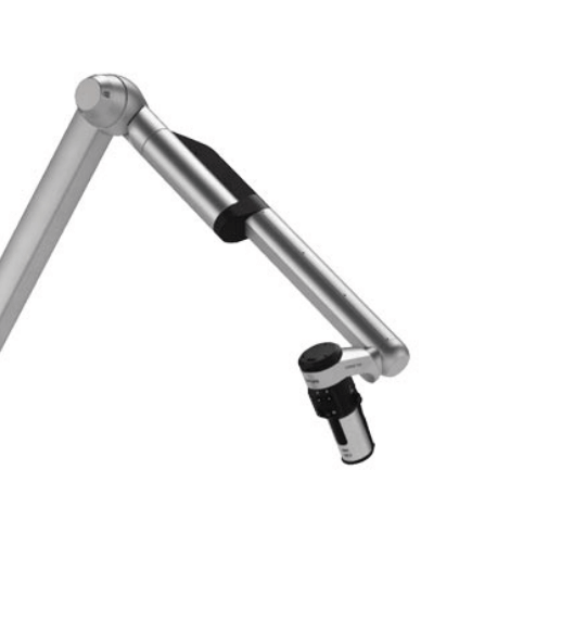
Over the last decade, spine surgery has undergone a revolution driven by ultra-high-resolution digital visualization. Following the era of the operating microscope (OM) and the advent of endoscopy, 4K–8K resolution 3D exoscopes emerge as the next major technological leap. These camera systems, mounted on an articulated arm, relay images to a stereoscopic monitor, freeing the surgeon from the “lens‑to‑eye” constraint and allowing the entire team to share the same surgical perspective.How does an exoscope work?
The exoscope combines large-format CMOS sensors, high-luminosity optics, and real-time image processing. The output is displayed on two monitors:
- Main 3D display (55–65″) positioned opposite the operating table.
- Auxiliary 2D/3D monitor for the rest of the team.
By projecting the scene in 3D, the system recreates the depth perception that classic microscopes relied on through ocular convergence. The surgeon controls zoom, focus, and viewing angle via foot pedals or a joystick, maintaining a working distance of up to 600 mm.
Evolution to 8K/3D
Early prototypes (2010–2015) provided 1080p 2D. In 2017, 4K 3D models were introduced, and since 2023 there have been 8K 3D platforms with 0.2 mm pixels at 40 cm. This upgrade effectively doubles the resolution lines of an operating microscope without compromising illumination or field of view. Comparative studies show that ultra-high definition identifies vasculo‑nervous structures under 100 µm with the same fidelity as a microscope, but with 40 % less optical readjustment time.
Clinical and ergonomic benefits
| Benefit | Reported impact | Evidence |
|---|---|---|
| Surgeon ergonomics | Reduction of cervical flexion angle from 42° to 12° | Innocenti et al. 2024 |
| Postoperative complications | ↓ Dural tears (1.8 % → 1.2 %) | ScienceDirect 2024 [2] |
| Surgical time | No difference vs. an operating microscope | JMISST 2025 [3] |
| Teaching | Shared field of view (100 % of the scene) | 2024 report [4] |
Key fact: In multicenter surveys, 87 % of neurosurgeons rate the exoscope’s ergonomics as “excellent,” compared to 23 % for operating microscopes.
Scientific evidence 2024–2025
- Prospective study of 67 minimally invasive TLIFs (Medicina, 2024): no differences in clinical outcomes; improved NASA‑TLX workload score.
- Systematic review of 54 articles (Cureus, 2025): the exoscope matches microscope safety and outperforms endoscopy in intradural surgeries.
- Narrative MISS‑Tech (JMISST, 2025): the exoscope + navigation reduces intraoperative radiation by 22 % [3].
- Comparative trial of 3D exoscope vs. operating microscope in lumbar discectomy (ScienceDirect, 2025): lower operator muscle fatigue (p < 0.01) and faster learning curve for residents [2].
Comparison with other visualization platforms
| Parameter | Operating microscope | Spinal endoscope | 8K/3D exoscope |
| Depth of field | Medium–high | Low | High |
| Angle of attack | Optically limited | Very limited | ±120° with an articulated arm |
| Team participation | Low (primary surgeon only) | Medium | High (entire OR sees the same image) |
| Ergonomics | Neck flexion >30° | Variable | Neutral posture |
From a cost–benefit standpoint, the exoscope falls between the operating microscope (more economical) and navigation robots (costlier). However, as an agnostic device – not tied to a single implant brand – its adoption does not create consumable dependencies.
Impact on various procedures
- Microtubular lumbar discectomy: enables a 16 mm incision, with return-to-work in 3.2 ± 0.8 weeks.
- Minimally invasive TLIF fusion: enhances contralateral visualization by avoiding tunnel shading.
- Intradural surgery: coaxial illumination reduces dural glare.
- Extramedullary cervical tumors: 4K/8K distinguishes tumor–cord interface in real time, aiding radiculomedullary blood supply preservation.
Patient experience
Various groups have incorporated postoperative 3D video in follow-up consultations. Patients understand the procedure better and request 28 % fewer clarification calls than with standard 2D video.
Standards and regulation
Exoscopes marketed in 2024–2025 comply with IEC 60601‑1 and IEC 60601‑2‑54 for surgical imaging equipment. Additionally, the FDA classifies them as Class II devices with partial 510(k) exemption when intended for neurosurgery, although 3D immersion validation reports are required.
Implementation strategies
- Pilot program: select a clinical champion and introduce the exoscope in microdiscectomies to familiarize the team.
- Training: simulation sessions with foam models or cadavers.
- Evaluation: NASA‑TLX metrics, technical error rates, and OR nurse satisfaction.
- Scaling: extend to scoliosis corrections, anterior cervical fusion, and complex intradural tumor procedures.
Integration with other technologies
- Intraoperative CT-guided navigation: semi-transparent overlay on the 3D image.
- Mixed reality and holography: headsets merging exoscope 3D output with anatomical models.
- Robotics: synchronized robotic arms maintain the visual axis while instrumentation robots place pedicle screws.
- 5-ALA fluorescence: selective visualization of intrathecal tumors without changing optics.
Limitations and challenges
- Initial cost (€300,000–€650,000) and maintenance contracts.
- Required 10–15 case learning curve to master hand-to-screen coordination.
- Low depth of field in very deep planes (solution: ND filters + automatic diaphragm).
- 3D visual fatigue in sensitive users; breaks every 90 minutes are recommended.
Preliminary economic analysis
An incremental cost study in three European university hospitals estimated a 3.4-year ROI for an 8K exoscope used in 50 % of spinal surgeries and 20 % of cranial cases. When considering:
- Reduced complications (−0.6 days of average stay; savings of €1,250 per episode).
- Increased surgeon productivity (−15 minutes of average anesthesia time, valued at €320).
- Higher training case volume (revenue from external rotations and workshops).
The authors conclude that even without technology-specific tariffs, early adoption can be sustainable for both public and private hospitals.
Required competencies
| Domain | Key skills | Recommended resources |
| 3D visualization | On-screen hand–eye coordination | VR microsurgery simulators |
| Arm control | Preset configuration and AXIS lock | Workshops with phantoms |
| Visual conditioning | Tolerance for prolonged stereoscopic viewing | Progressive stereopsis exercises |
| Data management | 4K/8K video capture and archiving | PACS with HEVC encoding |
Final conclusion
The 8K/3D exoscopy is emerging as the new reference standard for spinal surgery visualization. Its strategic adoption, backed by clinical evidence and a robust training program, promises to enhance patient safety and educational quality while optimizing the surgical team’s occupational health.
References
- Innocenti N, Corradino N, Restelli F, et al. High‑Definition 4K‑3D Exoscope in Spine Surgery: A Single‑Center Experience and Review of the Literature. Medicina (Kaunas). 2024;60(9):1476.
- ScienceDirect. Exoscope‑assisted spine surgery: Current applications and future perspectives. J Clin Orthop. 2024; in press.
- Alshaibi R, Mohamed AA, Williams C, et al. Exoscope Visualization, Navigation Guidance, and Robotic Precision in Spine Surgery. J Minim Invasive Spine Surg Tech. 2025;10(1):22‑33.
- Kalhorn CG. Changing the gold standard from operative microscope to 3D exoscope. Progressnotes – Institutional report. 2024.
- Levi V, Costa F. Surgeon experience with a digital exoscope: Multicenter survey. Neurosurg Rev. 2024;47(2):1023‑1034.
- Singh A, Kumar P, Dubey S, et al. Exoscope‑Assisted Spine Surgery: A Systematic Review From Basic to Complex Pathologies. Cureus. 2025;17.