Summary
Lumbar spinal canal stenosis is a narrowing that compresses the nerve roots and causes neurogenic claudication (pain or heaviness when walking that improves when sitting). Most people improve with conservative measures; when limitation persists, decompression (open, microtubular, or endoscopic) can help. In degenerative spondylolisthesis, 5-year data show that decompression without fusion offers comparable results to adding fusion in many cases. In 2025, trials support the non-inferiority of endoscopic decompression compared with microscopic decompression for selected indications.
- Key symptom: leg pain/numbness while walking that improves when sitting (neurogenic claudication).
- First, conservative: guided exercise, rational analgesia, and, in selected cases, injections.
- When to operate: persistent pain/disability after proper conservative management or progressive neurological deficit.
- Fusion: not automatic; in grade I spondylolisthesis, decompression alone is often enough.
- Emergencies: cauda equina syndrome = immediate hospital care.
- Recovery: without fusion, active life in 2–6 weeks; with fusion, 8–12+ weeks depending on job and comorbidities.
What is lumbar stenosis?
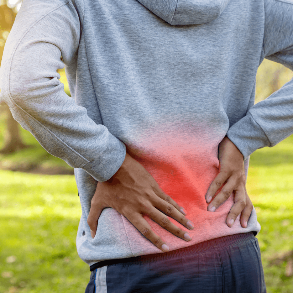
Lumbar spinal canal stenosis is the narrowing of the spinal canal or the lateral recesses/foramina due to degenerative changes (discs, facets, ligamentum flavum), which reduces the space for the nerve roots and causes pain/numbness, especially when standing or walking. It is more common after age 55–60 and may coexist with mild degenerative spondylolisthesis.
Symptoms and red flags
Typical presentation
- Pain/heaviness in buttocks and legs worsening with walking and improving when sitting or leaning forward.
- Tingling or weakness when walking uphill or standing for a long time.
- Low back pain may occur, but the main limiting factor is neurogenic claudication.
Red flags (urgent consultation or hospital)
- Progressive motor deficit in one or both legs.
- Cauda equina syndrome (CES): saddle anesthesia, urinary retention or incontinence, marked genital/perianal sensory changes.
- Fever or history of cancer (possible infection or metastasis).
Diagnosis: which tests are necessary (and which are not)
- History and examination guide the diagnosis.
- Lumbar MRI: test of choice if symptoms persist or diagnosis is uncertain; not routinely ordered for low back pain without neurological signs unless it will change management.
- Dynamic X-rays: if instability is suspected (e.g., spondylolisthesis).
- Electrophysiology: useful when neurological involvement or differential diagnoses are unclear.
Practical tip: MRI severity does not always match symptoms. Treat the person, not the image.
Step-by-step conservative treatment
- Education + exercise (flexion, endurance, and strengthening programs) and staying active — first-line.
- Pharmacologic: paracetamol/NSAIDs in short courses; avoid chronic opioids.
- Epidural/facet injections: temporary relief in selected cases; weigh risks/benefits.
- MILD (percutaneous decompression of the ligamentum flavum): minimally invasive alternative in some patients, with short- to mid-term safety/efficacy data; large long-term series are lacking.
Goal: improve walking capacity and quality of life, postponing or avoiding surgery if possible.
When to consider surgery
- Failure of well-executed conservative treatment (≥6–12 weeks) with significant functional limitation.
- Progressive neurological deficit.
- Severely impaired quality of life due to claudication despite adequate measures.
- Not decided based on MRI alone: the clinical correlation matters.
Surgical options: open, microtubular, and endoscopic
- Open decompression (laminectomy/laminotomy): classic standard, effective; more tissue disruption.
- Microtubular decompression: same concept with smaller incisions.
- Endoscopic decompression (uniportal or biportal): minimal incisions; trials and reviews show non-inferior functional results to microscopic decompression in selected patients, with less bleeding/stay in several series.
Limitations: learning curve and careful case selection.
Fusion: yes or no?
In grade I degenerative spondylolisthesis with stenosis, the NORDSTEN-DS trial (BMJ, 2024) showed that decompression alone is non-inferior to decompression + fusion at 5 years in disability (ODI) and reoperation rates. This does not mean fusion disappears: it may be indicated if there is clear instability, predominant axial mechanical pain, or demonstrated deformity/progression.
Real benefits and risks
Expected benefits
- Improvement in leg pain and walking capacity (main goal).
- Early discharge in minimally invasive/endoscopic techniques for selected cases.
Risks
- Dural tear (≈1–9%), infection, thrombosis, hematoma, reoperation for recurrence/adjacent segment disease.
- Fusion: adds risk of pseudarthrosis, greater bleeding and stay; no systematic superiority in grade I DS.
Recovery times and return to work
- Decompression without fusion: light active life in 2–6 weeks; return to work between 4–8 weeks (earlier in sedentary jobs).
- With fusion: return to work often 8–12+ weeks, depending on physical demands and recovery.
- Structured physiotherapy after surgery speeds up function and quality of life.
Key: occupation (sedentary vs physical) and preoperative condition affect return to work more than the technique itself.
When to go to the ER
- CES (saddle anesthesia, urinary retention/incontinence, marked genital/perianal sensory changes).
- Motor deficit worsening over hours–days.
- Fever and disproportionate pain with poor general condition.
AANS/NICE/ACR guidelines recommend urgent evaluation (MRI and early decompression if CES).
Myths and realities
- “Severe MRI = certain surgery.” Not always; clinical correlation comes first.
- “Fusion always improves outcomes.” Not in grade I DS according to 5-year RCTs.
- “Endoscopy = always better.” It is equivalent in selected indications; not all cases are suitable.
Patient checklist
- Does my walking pain improve when sitting?
- Have I tried exercise and rational analgesia for at least 6–12 weeks?
- Do I have progressive deficit or red flags? (if yes → ER)
- Is my case suitable for decompression without fusion? (assess stability)
- Which technique (open, micro, endoscopic) is most appropriate and why?
- What is my rehabilitation and return-to-work plan?
Frequently Asked Questions (FAQs)
Does lumbar stenosis always require surgery?
Many people improve with well-planned conservative care (exercise, rational analgesia, and selected injections).
When is surgery reasonable?
After ≥6–12 weeks of proper treatment without functional improvement or in case of progressive deficit.
Is fusion necessary if I have spondylolisthesis?
Not necessarily: in grade I, decompression alone may suffice (NORDSTEN-DS trial, 5 years).
Is endoscopic surgery as effective as open/microscopic?
In selected cases, RCTs and meta-analyses show non-inferior functional results and often less bleeding/stay.
When should I go to the ER?
If cauda equina syndrome appears: saddle anesthesia, urinary retention/incontinence, or genital/perianal sensory changes.
How long will it take to return to work?
Without fusion, 4–8 weeks (earlier if sedentary); with fusion, 8–12+ weeks.
Are injections useful?
They can provide temporary relief in selected cases; they do not correct the anatomical narrowing.
What are the risks of surgery?
Dural tear, infection, thrombosis, hematoma, and possible reoperation; with fusion, risk of pseudarthrosis and greater bleeding.
Glossary
Neurogenic claudication: Leg pain/heaviness when walking due to root compression, relieved by sitting.
- Decompression: Surgery to enlarge the nerve root space (laminectomy/laminotomy).
- Endoscopic (uni/biportal): Decompression using an endoscope with minimal incisions.
- Degenerative spondylolisthesis: Vertebral slippage due to arthritis.
- Fusion: Fixing two vertebrae with implants and graft to stabilize the segment.
- CES (cauda equina syndrome): Severe compression of terminal roots; surgical emergency.
References
- Dr. Vicenç Gilete – Neurosurgeon
- Dr. Augusto Covaro — Orthopedic and trauma surgeon
- Austevoll IM, et al. Decompression alone or with fusion for degenerative spondylolisthesis (NORDSTEN-DS): 5-year follow-up. BMJ. 2024. Link
- Park SM, et al. Biportal endoscopic vs microscopic decompressive laminectomy (RCT). Spine J. 2020. Link
- Kotheeranurak V, et al. Full-endoscopic vs tubular microscopic decompression (RCT). Eur Spine J. 2023. Link
- Chin BZ, et al. Endoscopic vs microscopic spinal decompression (systematic review). Spine J. 2024. Link
Disclaimer: This content is informational and does not replace the evaluation of a healthcare professional. If you suspect an emergency, go to the ER.