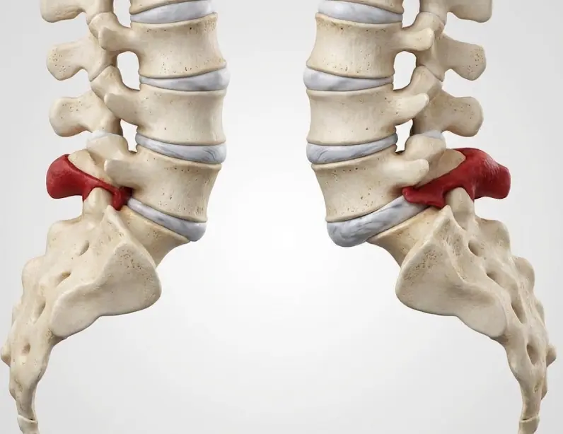
Lumbar spondylolisthesis is the “slipping” of one vertebra over the one below it. Sometimes it is an incidental finding with little relevance, and other times it is linked to sciatica, stenosis, and reduced walking tolerance. The decision is rarely an immediate “operate or don’t operate”: it is usually a stepwise process, with clear signals for when to escalate treatment.
- Many mild spondylolisthesis cases improve with a well-structured conservative plan over several weeks.
- Surgery is considered mainly when there is leg pain from nerve compression, persistent limitation, or progressive neurological deficit.
- In some cases, nerve decompression alone is enough; in others, stabilisation (fusion) is advisable if there is instability or predominantly mechanical pain.
- Recovery timelines depend more on the type of surgery (and your starting point) than on the diagnostic label itself.
What spondylolisthesis is (and why it is not always “serious”)
Spondylolisthesis occurs when one vertebra shifts forward relative to the one beneath it. In the lumbar spine, it most commonly affects L4-L5 or L5-S1. The slip may be small and stable (with few symptoms), or it may be associated with nerve compression, mechanical pain, and instability.
Most common types
- Degenerative: often appears with age due to wear of the disc and facet joints. It is frequently accompanied by stenosis (narrowing) of the spinal canal or the foramen where the nerve exits.
- Isthmic (pars defect or spondylolysis): more typical in active people or those with a history dating back to youth. The “pars” is a part of the vertebral arch that can develop a stress fracture from repetitive loading.
- Other, less common: dysplastic (congenital), traumatic, pathological (tumour/infection), or iatrogenic (after surgery).
How the “slip” is graded
It is usually classified by grade according to the percentage of forward translation. In low grades (I-II), many people live normally. In high grades, strategy and risks change, and specialist assessment is advisable.
Symptoms and indications: when to worry and which pattern matters
The diagnosis alone does not determine treatment. What matters is the combination of symptoms, examination, and tests.
Common symptoms
- Mechanical low back pain: worse when standing, with extension (leaning the trunk backwards), or when lifting; improves when sitting or lying down.
- Sciatica or radiculopathy: pain radiating through the buttock and down the leg, with tingling, cramps, or weakness depending on the affected nerve root.
- Neurogenic claudication: “heavy” legs or pain while walking that improves with sitting or bending forward. This often suggests associated stenosis.
Priority red flags (not a “wait and see” situation)
- New or progressive weakness in one or both legs (for example, a “foot drop”).
- Bowel or bladder changes (urinary retention, incontinence) or saddle anaesthesia (genital/perineal numbness).
- Pain with fever, chills, or feeling generally unwell (possible infection).
- History of cancer plus persistent night pain or unexplained weight loss.
Diagnosis: which tests are usually useful (and what question each one answers)
The goal of the work-up is to answer four questions: (1) where the pain is coming from (nerve, disc, facets, sacroiliac joint), (2) whether there is neurological compression, (3) whether instability is present, and (4) whether there are factors that change the strategy (fragile bone, deformity, prior surgery).
Clinical examination
- Strength, reflexes, and sensation in the legs.
- Neural tension tests (for example, straight-leg raise).
- Hip and sacroiliac assessment to avoid confusing pain sources.
Most common imaging tests
- Standing X-rays: show the slip under load and help assess alignment.
- Dynamic X-rays (flexion-extension): can help detect instability in some cases (not all).
- MRI: evaluates nerves, stenosis, discs, and soft tissues.
- CT: useful if a pars defect is suspected, to visualise bone, or when prior surgery requires more bony detail.
When “extra” tests are typically requested
- Bone density scan and bone labs if osteopenia/osteoporosis is suspected and surgery is being considered.
- Diagnostic blocks (in selected cases) when it is unclear whether pain is facet-related, sacroiliac, or from another source.
Step-by-step conservative treatment (what helps most when done well)
In mild to moderate spondylolisthesis without progressive deficit, it is common to start with conservative measures for several weeks. “Conservative” does not mean giving up: it means an active plan that is measurable and adjustable.
1) Education and load management
- Learn which movements trigger symptoms (often sustained extension and loading) and how to dose them without stopping movement altogether.
- Avoid prolonged rest: it usually worsens stiffness and reduces exercise tolerance.
2) Therapeutic exercise and physiotherapy
- Lumbopelvic control and core: improve functional stability.
- Glutes and posterior chain: strength and endurance for walking and standing.
- Hip mobility: can sometimes reduce lumbar overload.
- Gait training if there is neurogenic claudication (with gradual progression).
3) Thoughtful pain relief and complementary strategies
Medication, if used, should be reviewed by a professional (especially with comorbidities). In some profiles, injections are considered to relieve radicular pain or to refine the diagnosis. The key is that these measures support the active plan rather than replace it.
When to conclude conservative care is “not working”
- Leg pain or walking limitation remains high despite adherence to an active plan.
- Neurological worsening.
- Meaningful functional impact (work, sleep, self-care) that does not improve.
Surgical options: decompression and fusion (and why you may not always need “fixation”)
Surgery is not a punishment or a failure: it is a tool to consider when the benefit-risk balance is favourable. In spondylolisthesis, the two main surgical families are:
Option A: Decompression (freeing the nerve)
If the main issue is nerve compression (sciatica, neurogenic claudication) due to associated stenosis, decompression aims to create space for the nerve root or the canal. It can be performed with more or less invasive techniques depending on the case. The goal is to reduce leg pain and improve walking capacity.
Option B: Fusion (stabilisation)
Fusion (arthrodesis) joins two vertebrae to eliminate painful or unstable motion. It is typically considered when there are clear signs of meaningful instability, deformity, predominantly axial mechanical pain that matches the level, or when a wide decompression could leave the segment too “loose.”
The big question: decompression alone or decompression plus fusion?
In low-grade degenerative spondylolisthesis, recent trials and reviews have shown that, in selected patients, decompression alone can provide comparable outcomes in function and pain to adding fusion, with less surgical burden. This does not mean fusion is “obsolete”: it means it should not be automatic. The decision depends on stability, pain pattern, anatomy, and functional goals.
What tips the balance one way or the other
- More in favour of decompression alone: symptoms dominated by leg pain/claudication, low and stable slip, little axial mechanical pain, and no significant deformity.
- More in favour of adding stabilisation: clear instability on dynamic films, predominant and concordant mechanical back pain, documented progression, deformity, or need for a decompression that compromises stability.
Real-world benefits and risks (no promises)
Expected benefits
- If leg pain predominates: improvement typically focuses on sciatica and walking.
- If mechanical pain with instability predominates: well-indicated stabilisation can improve pain and standing tolerance.
Risks and adverse effects to understand
- Infection, bleeding, thrombosis, anaesthetic complications.
- Nerve injury or irritation (temporary or, rarely, persistent).
- CSF leak from a dural tear (more likely in revisions or complex stenosis).
- If fusion is performed: non-union (pseudoarthrosis), implant failure, and potential adjacent-level problems over time.
- Possibility of reoperation: depends on the case, technique, bone biology, and clinical course.
When to seek referral and a second opinion (practical criteria)
- Persistent leg pain with neurological signs (strong tingling, loss of strength, abnormal reflexes).
- Neurogenic claudication that limits walking despite a structured conservative plan.
- Uncertainty about instability or about the need for fusion.
- History of prior surgery, osteoporosis, deformity, or multiple affected levels.
- Need to understand options with realistic expectations (likely benefit, risks, timelines, restrictions).
Recovery: realistic timelines (typical ranges, and why they vary so much)
Timelines vary depending on your starting point (pain, strength, fitness), the type of surgery, and the demands of your job. Still, these ranges are often useful for orientation:
If there is decompression without fusion
- Walking: early (often the same day or the next day).
- Light activities at home: first few days.
- Sedentary work: often between 2 and 6 weeks, if pain is controlled and you can take breaks.
- Strain and lifting: gradual progression over 6 to 12 weeks, depending on recovery.
If there is fusion (stabilisation)
- Functional recovery is usually measured in months.
- Sedentary work: often between 6 and 12 weeks (highly variable).
- Physical work: often 3 to 6 months or more, depending on the fusion type and your progress.
- Bone consolidation: can take several months; habits (smoking), nutrition, and bone health matter.
Key idea: the goal is not to be “perfect” by a fixed date, but to restore function progressively and safely, avoiding load spikes that trigger setbacks.
When to go to emergency care
- Urinary retention, incontinence, or numbness in the genital/perineal area.
- Rapid or progressive weakness in one or both legs.
- High fever with severe low back pain or sudden worsening of pain with feeling unwell.
- After surgery: wound drainage, marked redness, uncontrolled pain, shortness of breath, or chest pain.
Myths and realities
Myth: “If I have spondylolisthesis, I will end up needing surgery”
Reality: many low-grade slips are managed without surgery, especially if there is no progressive deficit and the conservative plan is well designed.
Myth: “The MRI decides the treatment”
Reality: imaging helps, but the decision depends on symptoms, examination, function, and progression. Some MRIs look “dramatic” with few symptoms, and the reverse also happens.
Myth: “Fusion is always better”
Reality: in some profiles, adding fusion provides stability and improves outcomes; in others, it may not add clear benefit and does add surgical burden. Patient selection is the key.
FAQs
Does spondylolisthesis always hurt?
No. It can be an incidental finding on an X-ray done for another reason. When it causes symptoms, they are usually related to mechanical low back pain, nerve compression (sciatica), or stenosis with reduced walking tolerance.
Can it be “corrected” with exercises?
Exercises do not usually move the vertebra “back into place,” but they can reduce pain and improve function by increasing functional stability, strength, and load tolerance. In many cases, that is enough to live well.
What does it mean if it is degenerative?
It means it is associated with progressive wear of discs and joints with age. It more often coexists with stenosis and walking-related symptoms than with sudden acute pain after a single effort.
What is the difference between spondylolysis and spondylolisthesis?
Spondylolysis is a stress defect or crack in part of the vertebral arch (the pars). It can exist without slippage. Spondylolisthesis is the slippage; it sometimes occurs on top of pre-existing spondylolysis (isthmic type).
When is surgery considered?
Mainly when there is leg pain from nerve compression or neurogenic claudication that does not improve with a well-executed conservative plan, or when progressive weakness appears. The surgical goal is usually to free the nerve and, if needed, stabilise.
Do you always need screws (fusion)?
Not always. In low-grade degenerative spondylolisthesis, some people improve with decompression without fusion. In others, because of instability or predominantly mechanical pain, stabilisation may be more reasonable. It is decided case by case.
How long does recovery take?
It depends on the type of surgery and your job. After decompression without fusion, many people return to sedentary tasks in a few weeks. After fusion, recovery is usually longer and measured in months.
What should I do if I have tingling or loss of strength?
If it is new, intense, or progressing, prompt medical evaluation is advisable. If it comes with urinary problems or perineal numbness, it is an emergency.
Glossary
- Spondylolisthesis
- Slippage of one vertebra over another.
- Spondylolysis (pars defect)
- Stress fracture or defect in part of the vertebral arch (pars interarticularis).
- Radiculopathy
- Symptoms from irritation or compression of a nerve root (radiating pain, tingling, weakness).
- Stenosis
- Narrowing of the canal or foramen that reduces space for nerves.
- Decompression
- Surgery to free compressed nerves (for example, removing tissue that narrows the canal).
- Fusion (arthrodesis)
- Surgery that joins vertebrae to stabilise and eliminate painful or unstable motion.
- Pseudoarthrosis
- Failure of bone to fuse after a fusion procedure, with possible pain or mechanical failure.
- Neurogenic claudication
- Leg pain or heaviness while walking due to nerve compression, improving with sitting.
Health education disclaimer: this content is informational and does not replace an individual medical assessment. If you have neurological symptoms, progressive worsening, or red flags, consult healthcare professionals or seek emergency care.
References and clinical guidelines
- North American Spine Society (NASS).
Diagnosis and Treatment of Degenerative Lumbar Spondylolisthesis. Clinical guideline (PDF).
Year: 2014. Link to the document
- North American Spine Society (NASS).
Degenerative Lumbar Spondylolisthesis Appropriate Use Criteria. PDF.
Year: 2021.Link to the document
- Austevoll IM, et al.
Decompression with or without Fusion in Degenerative Lumbar Spondylolisthesis.
New England Journal of Medicine.
Year: 2021.Link to the article
- Gadjradj PS, et al.
Systematic review on routine fusion in degenerative spondylolisthesis.
EClinicalMedicine.
Year: 2022. Link to the article