Endoscopic spine surgery decompresses nerves through tiny incisions, minimizing soft-tissue injury and shortening hospital stays. When properly indicated, it offers pain relief and functional improvement comparable to classic microdiscectomy. Not every case is a candidate, and risks and recurrences still exist; individualized selection and team experience are decisive.
- Best suited for lumbar/cervical disc herniation and select cases of foraminal stenosis.
- Pain and function outcomes comparable to microdiscectomy; hospital stay and recovery are often shorter.
- Does not eliminate risks: infection, nerve injury, herniation recurrence, CSF leak.
- Decision follows clinical–radiologic correlation and failure of appropriate conservative treatment.
What is endoscopic spine surgery?
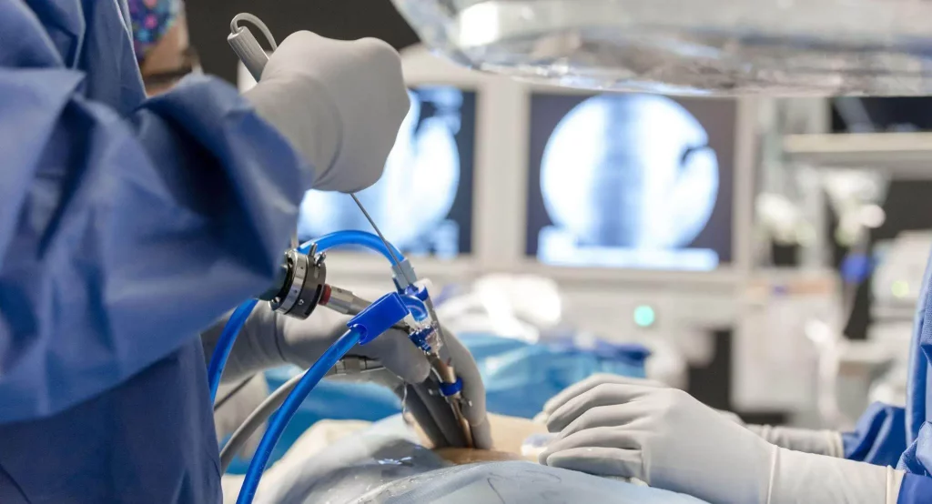
A set of minimally invasive techniques that use a camera and microinstruments introduced through one or two portals to reach the disc or spinal canal. The most common variants are the transforaminal endoscopic approach (TELD) and uniportal or biportal endoscopy. The goal is to remove the fragment compressing the nerve root or enlarge the nerve corridor while preserving as much healthy tissue as possible.
Who might be a candidate? Symptoms and indications
The most frequent indication is disc herniation with radiculopathy (sciatica or neck pain radiating to the arm) that does not improve after a well-structured conservative plan for 6–8 weeks. It may also be considered in:
- Foraminal or lateral stenosis with pain on walking or standing.
- Recurrent disc herniation in selected patients.
- Some focal compressions after previous surgery, when anatomy allows.
Red flags requiring urgent evaluation: progressive weakness, saddle anesthesia, loss of bladder or bowel control, or sudden, disabling pain.
Diagnosis: confirming whether endoscopy is appropriate
The decision is based on correlating symptoms, neurological exam, and imaging:
- MRI to locate the conflict (disc fragment, foraminal/canal stenosis).
- Dynamic X-rays to rule out clinically relevant instability.
- CT useful in recurrences or prior surgeries to assess pars, facets, and implants.
- Level selection and planning a safe corridor (for example, Kambin’s triangle in transforaminal approaches).
Treatment alternatives
Non-surgical
- Pain education and therapeutic exercise (walking, core strengthening).
- Prudent pharmacologic therapy (non-opioid analgesics; judicious use of adjuvants).
- Active physiotherapy and posture measures.
- Injections or radiofrequency in selected cases (e.g., facet/sacroiliac pain after ruling out other causes).
Surgical
- Microdiscectomy (traditional gold standard) with proven long-term results.
- Microtubular decompression and laminotomy for central or combined stenosis.
- Fusion when there is significant instability or deformity requiring it.
Expected benefits and existing risks
Recent comparative reviews show that endoscopic discectomy achieves pain relief and functional improvement similar to microdiscectomy. Common advantages include less blood loss, less postoperative pain, lower opioid use, and shorter hospital stays. Across multiple series and meta-analyses, overall complication rates are comparable, with some reviews suggesting even lower figures for endoscopy. However, risks remain: infection, hematoma, nerve injury, dysesthesia from root irritation, cerebrospinal fluid leak, thrombosis, wrong level in settings without navigation, and recurrent herniation (especially in discs with a weakened annulus).
The learning curve and case selection influence outcomes: highly migrated herniations, multilevel central stenosis, or marked instability may require other techniques.
Practical referral criteria
- Disabling radiculopathy with imaging correlation after 6–8 weeks of appropriate management.
- Progressive neurological deficit or pain preventing basic independence.
- Recurrent herniation with good clinical correlation and a safe window for endoscopic access.
- Focal foraminal/lateral stenosis when anatomy favors a targeted decompression.
Realistic recovery timelines
- First 24–48 h: early ambulation, multimodal analgesia, discharge often same day or within 24 h if uneventful.
- Week 1–2: daily walks; return to light office tasks if pain allows.
- Week 3–6: gradual strength and endurance work; many patients resume jobs with low physical demand.
- 6–12 weeks: graded return to demanding physical activity; impact sports only with clearance.
These time frames are approximate and depend on age, comorbidities, hernia/stenosis type, treated level, and job demands.
When to seek emergency care?
- Sudden or progressive weakness in foot, hands, or legs.
- Loss of bladder/bowel control or saddle anesthesia.
- High fever with severe pain after surgery.
- Progressive, disabling pain not controlled with scheduled analgesia.
Myths and facts
- Myth: “Endoscopy always heals faster than any other technique.” Fact: recovery is often quicker, but outcomes depend on diagnosis and case selection.
- Myth: “It’s a minor surgery without risks.” Fact: it reduces tissue trauma but does not eliminate complications.
- Myth: “If I have endoscopic surgery, I won’t get another herniation.” Fact: recurrence is possible; habits, load, and anatomy matter.
Frequently asked questions
How does it differ from “classic” microdiscectomy?
Both aim to decompress the nerve. Endoscopy uses portals and irrigation; microdiscectomy uses a larger incision and a microscope. Pain and function outcomes are comparable; endoscopy often shortens hospital stay.
Is it performed under general anesthesia?
It can be done under general or regional/epidural anesthesia in experienced centers. The choice depends on approach, level, and comorbidities.
Is it suitable for extensive “central canal” stenosis?
Not always. In multilevel or severe central stenosis, microtubular or open decompression may be preferable.
Can I return to work soon?
Office work: often within 1–3 weeks if recovery is smooth. Manual work: 6–12 weeks with progressive reconditioning.
How long does the procedure take?
About 45–120 minutes depending on level, hernia type, and access route.
What are the chances of reoperation?
Similar to microdiscectomy in large series. Recurrent herniation, when it occurs, usually appears in the first months.
Glossary
- TELD: transforaminal endoscopic lumbar discectomy.
- UBED: unilateral biportal endoscopy (two portals).
- Radiculopathy: pain/changes due to irritation of a nerve root.
- Laminotomy: partial opening of the posterior arch to decompress.
- CSF leak: cerebrospinal fluid leakage due to dural tear.
References
- Complex Spine Institute.
https://complexspineinstitute.com/en/complex-spine-institute/en/treatments/endoscopic-spine-surgery/ - BMJ. Full endoscopic versus open discectomy for sciatica (meta-analysis). 2022. https://www.bmj.com/content/376/bmj-2021-065846
- NICE NG59. Low back pain and sciatica in over 16s: assessment and management (2020 update). 2020. https://www.nice.org.uk/guidance/ng59
- Neurospine. TELD vs microdiscectomy: follow-up >5 years. 2024. https://www.e-neurospine.org/journal/view.php?number=1535
- PubMed. UBED vs microdiscectomy in lumbar herniation. 2024. https://pubmed.ncbi.nlm.nih.gov/38388729/
- PLOS/PMC. Microdiscectomy, IPD and PELD: similar clinical outcomes. 2025. https://pmc.ncbi.nlm.nih.gov/articles/PMC11942998/
- NASS. Clinical Guidelines (official resource). 2021–2025. https://www.spine.org/Research/Clinical-Guidelines
Disclaimer: This content is educational and does not replace individual medical assessment or advice from a healthcare professional.