- Not every neck pain or dizziness is “instability”: diagnosis requires symptoms + examination + imaging guided by a clear clinical question.
- Two related concepts are often discussed: craniocervical instability (skull-C1) and atlantoaxial instability (C1-C2). They are sometimes grouped as “upper cervical instability.”
- No radiologic measurement alone confirms the diagnosis: numbers must be interpreted in context.
- Treatment usually starts with well-targeted non-surgical options; a cervical collar can help as a short trial in specific cases, but it is not a long-term solution.
- Surgery (instrumentation/fusion) is reserved for selected situations, with clinical and radiologic criteria and a clear benefit-risk balance.
- If there is progressive weakness, gait change, loss of bladder or bowel control, or breathing difficulty, do not wait: urgent assessment is needed.
What it is and why it’s so often confused
The craniocervical junction is a “hinge” region formed by the occipital bone (base of the skull) and the first two cervical vertebrae: the atlas (C1) and the axis (C2). It is designed to allow movement while protecting critical structures: the upper spinal cord, brainstem, lower cranial nerves, and vertebral arteries.
We talk about craniocervical instability when, due to ligament laxity, structural changes, or sequelae of trauma/inflammation, that hinge moves abnormally or is controlled less effectively, and this correlates with symptoms. If the main problem is between C1 and C2, it is often called atlantoaxial instability. Many sources group these as upper cervical instability.
Why so much confusion? Because occipital pain, dizziness, brain fog, fatigue, or palpitations can have many causes. In addition, some people with hypermobility or connective tissue disorders have complex symptoms and comorbidities (for example, migraine, dysautonomia), which makes it easy to attribute everything to a single explanation. The key is to go step by step: safety first, then a reasoned diagnosis.
9 signs that justify looking into it (and 6 that often mislead)
9 signs that do deserve a structured evaluation
- Occipital or suboccipital pain (at the back of the head near the skull base) that worsens with sustained postures or specific movements, and partially improves with cervical support.
- A “heavy head” sensation or needing to support the head with the hands by the end of the day.
- Neurological symptoms consistent with upper spinal cord involvement: hand clumsiness, gait disturbance, stiffness or “odd legs,” unexplained falls.
- New or progressive swallowing or voice problems (without an obvious ENT cause), especially if they occur alongside other neurological signs.
- Repeated syncope or near-syncope that seems related to neck positions (note: this can also occur in dysautonomia, which is why careful evaluation matters).
- Pain and symptoms after significant neck trauma (for example, whiplash) with a difficult course despite appropriate rehabilitation.
- Clear worsening with neck flexion/extension (beyond typical mechanical pain), with “systemic” or neurological symptoms.
- A history of significant hypermobility with disabling cervical symptoms, especially if there are signs of instability in other joints.
- Imaging findings compatible with altered upper cervical alignment and clinical correlation (findings without symptoms are not enough).
6 things that often mislead (and don’t prove anything on their own)
- Clicks or “cracking” in the neck without neurological symptoms: common and often benign.
- Nonspecific dizziness without a clear pattern: it may be vestibular, migrainous, anxiety-related, medication-related, or autonomic.
- Chronic headache: migraine and tension-type headache are far more common.
- An MRI “showing disc bulges” in the neck: very common even in people without pain.
- Feeling better with a collar for months: this may reflect postural dependence or muscular “rest,” not a reliable diagnostic test.
- Isolated radiologic measurements interpreted out of context: numbers without a clinical exam can lead to overdiagnosis.
Diagnosis: which tests truly help
A useful diagnosis starts with three questions: 1) are there red flags or neurological injury?, 2) do more common causes fit better?, 3) if we suspect instability, which test will change decisions?
Clinical examination (the part you can’t skip)
The goal is to look for signs of myelopathy (hyperreflexia, gait changes, clumsiness), lower cranial nerve deficits (swallowing/voice), radicular signs, and postural tolerance. History matters too: trauma, prior surgery, systemic inflammation, hypermobility, and which treatments have already been tried.
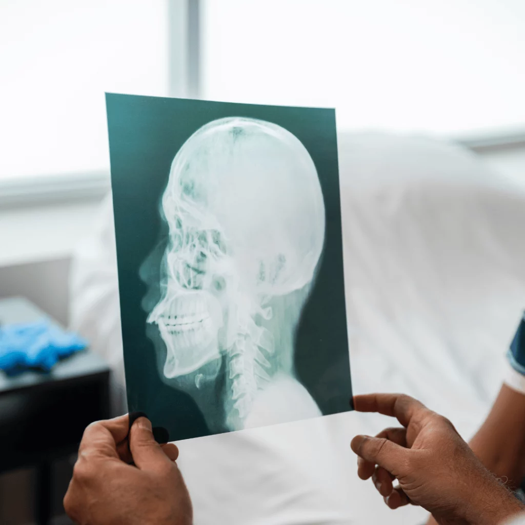
Imaging: what each test usually adds
- Dynamic X-rays (flexion/extension): help identify abnormal motion and alignment under functional loading. Useful when mechanical instability is suspected.
- MRI: evaluates the spinal cord, upper brainstem, discs, and partly the ligaments. Essential if there are neurological signs.
- CT scan: shows bony anatomy in detail and may help when bone anomalies or sequelae are suspected, or for surgical planning.
- Seated or standing studies (when available): may provide additional information about alignment under load in some cases. Not necessary for everyone.
Measurements you’ll see online (and how to understand them without falling into traps)
In the literature you’ll find metrics such as the clivo-axial angle, Harris-type measurements, or the Grabb-Mapstone-Oakes line. They describe the skull-C1-C2 relationship and potential deformation/angulation. The key point is this: there is no single “magic number.” Normal ranges exist, techniques vary, and rigid cutoffs without clinical correlation increase the risk of overdiagnosis.
Differential diagnosis: common things first
Before labeling “instability,” it’s important to rule out common, treatable causes: migraine (including vestibular migraine), temporomandibular disorders, peripheral vestibular disease, sleep disorders, anemia/thyroid problems, medication effects, degenerative cervical stenosis/compression, and dysautonomia. Sometimes conditions coexist: the goal is not to pick a single label, but to explain the picture and prioritize risk.
Non-surgical options and when they’re considered
In many cases, even with hypermobility, the first step is conservative care. This doesn’t mean “just endure it”: it means treating methodically, measuring response, and adjusting.
Education and load management
- Identify triggering postures (phone use, laptop work, driving, looking upward).
- Short, frequent breaks tend to work better than “total rest.”
- Avoid extreme cervical ranges if they provoke symptoms, but don’t immobilize yourself long-term.
Targeted physical therapy (not “just massage”)
The aim is usually better motor control and tolerance: deep neck flexors, cervical stabilization, scapular girdle, breathing, and proprioception. With hypermobility, progression is slower and pushing into end ranges is avoided. If a technique clearly and persistently worsens symptoms, the plan should be reconsidered.
Cervical collar: helpful as a short trial, risky as a long-term crutch
A rigid or semi-rigid collar can be used briefly to see whether reducing motion changes symptoms, or for specific situations. If used for weeks or months without an exit strategy, it can cause muscular deconditioning and dependence. If considered, it should come with a plan (when, how long, how to taper off).
Pain and comorbidity management
Depending on the case: cautious analgesia, migraine treatment, vestibular therapy, dysautonomia management (hydration, salt if appropriate, compression stockings, graded routines), sleep hygiene, and psychological support if there is pain sensitization. It’s not “all psychological”: mechanical and nervous system factors influence each other.
Surgical options: benefits, risks, and limits
Surgery is not the “definitive test,” and it’s not a shortcut. It is reserved for selected cases with a consistent clinical pattern, radiologic correlation, and a favorable benefit-risk balance.
Which surgeries are considered
- Occipitocervical fusion (skull to upper cervical spine) when the main problem is at the skull-C1 junction, or when there is neurological compromise and pathologic alignment.
- C1-C2 fusion (atlantoaxial) when the dominant instability is between atlas and axis.
- In specific contexts, it may be combined with decompression if there is significant neural compression from another cause.
Expected benefits (no promises)
In selected series, improvements are reported in head/neck pain, neurological symptoms, and quality of life. But the evidence is heterogeneous and partly observational, which is why patient selection is critical and a discussion of expectations is essential.
Risks and adverse effects (what you should understand before deciding)
- Loss of upper cervical mobility (rotation and flexion-extension) to a greater or lesser degree depending on the fused levels.
- Surgical complications: infection, bleeding, nerve or vascular injury, wound problems, thrombosis.
- Mechanical complications: hardware failure, pseudarthrosis, adjacent-level issues over the medium to long term.
- Persistent symptoms: if part of the picture comes from migraine/dysautonomia or other causes, fusion won’t “fix” everything.
When to refer, realistic recovery timelines, and emergencies
Practical referral criteria (priority consultation)
- Neurological symptoms suggestive of myelopathy (gait changes, clumsy hands, falls) even if intermittent.
- Severe, limiting occipital pain with a clear postural component and poor response to a well-executed conservative plan.
- Major hypermobility history with disabling cervical symptoms and a reasoned suspicion of instability.
- Imaging findings that could justify decisions (and not just incidental findings).
Realistic recovery timelines (approximate)
With conservative management: usually measured in weeks to months. A “reliable” change is often seen after 6 to 12 weeks of a well-executed plan, with adjustments.
After fusion surgery: early functional recovery is often weeks, but consolidation and adaptation are measured in months.
- Hospital stay: often 2 to 5 days, depending on complexity and comorbidities.
- First 2 to 6 weeks: pain control, safe mobility, start of prescribed rehabilitation.
- 6 to 12 weeks: progressive activity increase; some desk-based jobs can be resumed gradually if recovery is going well.
- 3 to 6 months: improved endurance and stability; medication and physical therapy adjustments.
- 6 to 12 months: bone fusion and final outcome assessment in many cases.
When to go to the emergency department
- New or progressive weakness, marked loss of strength, difficulty walking, or falls.
- Loss of bladder or bowel control or numbness in the perineal area.
- Breathing difficulty, significant choking, or sudden worsening of swallowing.
- Sudden, unusual headache (“the worst”), fever with marked stiffness, or feeling very unwell.
Myths and realities
- Myth: “If I have dizziness and neck pain, it must be instability.” Reality: much more common causes exist; the key is an orderly differential diagnosis.
- Myth: “One imaging measurement proves it.” Reality: numbers must be interpreted with symptoms, examination, and technique quality.
- Myth: “A collar confirms the diagnosis.” Reality: it can guide, but it can also mislead due to postural unloading effects.
- Myth: “Surgery fixes everything.” Reality: it may improve a mechanical-neurological component, but it doesn’t erase comorbidities or pain sensitization.
Frequently asked questions
Is craniocervical instability common?
No. In neck pain clinics, the most common causes are degenerative, muscular, or migraine-related. Upper cervical instability exists, but it is less common and requires clear criteria to avoid overdiagnosis.
Can it exist with a “normal” supine MRI?
In some cases, alignment under load or during motion may add information. Even so, a normal MRI does not rule out all causes, and an MRI with findings does not confirm that those findings explain your symptoms. Context is decisive.
Is a cervical collar a diagnostic test?
It can be an orienting tool if used briefly with a defined goal (for example, seeing whether restricting motion changes a symptom). It is not definitive, and prolonged use can worsen things through deconditioning.
If I have hypermobility or Ehlers-Danlos, does that mean I have upper cervical instability?
Not necessarily. Hypermobility increases the risk of joint control problems, but most people do not have severe instability. What matters is the combination of symptoms, examination, and appropriately indicated tests.
Can physical therapy make me worse?
If end-range is forced, aggressive manipulation is used, or progression is too fast, yes, it can worsen symptoms. Helpful therapy is usually specific, gradual, and focused on motor control while avoiding extremes. If a plan persistently worsens you, it should be adjusted.
Is high-velocity cervical manipulation advisable if instability is suspected?
In general, it is not a first recommendation when upper cervical instability or neurological symptoms are suspected. Safety, diagnosis, and stabilization/control strategies come first.
What outcomes can I expect if surgery is ultimately recommended?
It depends on the reason for surgery, the clinical-radiologic correlation, and comorbidities. In selected patients it can improve pain and certain neurological symptoms, but there is loss of mobility and risk. The decision must be highly individualized.
How long does it take to notice improvement with conservative treatment?
If the plan is well indicated and followed, some people notice changes in 2 to 4 weeks, but stable improvement usually requires 6 to 12 weeks, and sometimes longer.
Glossary
- Craniocervical instability
- Abnormal movement or control at the junction between the skull and C1 that correlates with symptoms and, in some cases, neurological involvement.
- Atlantoaxial instability
- Instability that predominates between C1 (atlas) and C2 (axis).
- Myelopathy
- Spinal cord dysfunction. It can cause clumsiness, gait disturbance, hyperreflexia, or weakness.
- Clivo-axial angle
- A radiologic measurement describing the relationship between the skull base and the cervical axis; it must be interpreted in context.
- Pseudarthrosis
- Lack of bony healing after a fusion.
- Dysautonomia
- Dysfunction of the autonomic nervous system (for example, orthostatic intolerance, palpitations, dizziness).
References
- Craniocervical Instability in Ehlers-Danlos Syndrome: A Systematic Review (2022)
- Craniocervical instability in patients with Ehlers-Danlos syndromes: outcomes analysis following occipito-cervical fusion (2024)
- Presentation and physical therapy management of upper cervical instability in symptomatic generalized joint hypermobility (2023)
- Non-surgical assessment and management of upper cervical instability (UCI) (2023)
- Reference values of measures of craniocervical junction alignment (warning about over-diagnosis with rigid cutoffs) (2023)
- Normal range of clivo-axial angle in adults using flexion and extension MRI (2021)
- Association between clivo-axial angle and distal junctional failure after craniocervical fusion (2025)
- Co-occurrence of tethered cord syndrome and cervical spine instability in hypermobile EDS (2024)
- Comorbidities and neurosurgical interventions in a cohort with hypermobile EDS (2025)
- Impact of imaging modality on craniocervical junction angles (2019)
Health notice: This content is educational and does not replace individual medical assessment. If you have neurological symptoms, rapid worsening, or red flags, seek a qualified clinician or go to emergency care.